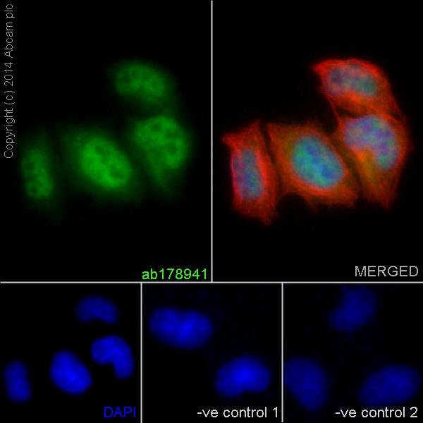
Immunofluorescent analysis of 4% paraformaldehyde-fixed HeLa (Human epithelial cells from cervix adenocarcinoma) cells labeling STAT5b with ab178941 at 1/100 dilution. The cells were permeabilised with 0.1% Triton X-100. Goat anti-rabbit IgG (Alexa Fluor® 488) (ab150077) at 1/200 dilution was used as the secondary antibody (green). Nuclear and cytoplasm staining is detected. The nuclear counter stain is DAPI (blue). Tubulin is detected with ab7291 (Tubulin mouse mAb) at 1/500 and ab150120 (AlexaFluor®594 Goat anti-Mouse secondary) at 1/400 dilution (red). The negative controls are as follows;1. ab178941 at 1/100 dilution followed by Goat anti mouse IgG (Alexa Fluor®594) at 1/400 dilution.2. ab7291 (anti-Tubulin mouse mAb) at 1/500 dilution followed by Goat anti rabbit IgG (Alexa Fluor®488) ar 1/200 dilution.
![All lanes : Anti-STAT5b [EPR16671] antibody (ab178941) at 1/20000 dilutionLane 1 : K562 (Human chronic myelogenous leukemia cells from bone marrow) whole cell lysatesLane 2 : HeLa (Human epithelial cells from cervix adenocarcinoma) whole cell lysatesLane 3 : Jurkat (Human T cell leukemia cells from peripheral blood) whole cell lysatesLane 4 : Daudi (Human Burkitt's lymphoma cell line) whole cell lysatesLysates/proteins at 20 µg per lane.SecondaryGoat Anti-Rabbit IgG, (H+L), Peroxidase conjugated at 1/1000 dilution](http://www.bioprodhub.com/system/product_images/ab_products/2/sub_5/5875_ab178941-228444-178941.jpg)
All lanes : Anti-STAT5b [EPR16671] antibody (ab178941) at 1/20000 dilutionLane 1 : K562 (Human chronic myelogenous leukemia cells from bone marrow) whole cell lysatesLane 2 : HeLa (Human epithelial cells from cervix adenocarcinoma) whole cell lysatesLane 3 : Jurkat (Human T cell leukemia cells from peripheral blood) whole cell lysatesLane 4 : Daudi (Human Burkitt's lymphoma cell line) whole cell lysatesLysates/proteins at 20 µg per lane.SecondaryGoat Anti-Rabbit IgG, (H+L), Peroxidase conjugated at 1/1000 dilution
![All lanes : Anti-STAT5b [EPR16671] antibody (ab178941) at 1/5000 dilutionLane 1 : Mouse brain lysatesLane 2 : Mouse heart lysatesLane 3 : Mouse kidney lysatesLane 4 : Mouse spleen lysatesLane 5 : Rat brain lysatesLane 6 : Rat heart lysatesLane 7 : Rat kidney lysatesLane 8 : Rat spleen lysatesLane 9 : C6 (Rat glial tumor cells) whole cell lysatesLane 10 : RAW 264.7 (Mouse macrophage cells transformed with Abelson murine leukemia virus) whole cell lysatesLane 11 : PC-12 (Rat adrenal gland pheochromocytoma) whole cell lysatesLane 12 : NIH/3T3 (Mouse embyro fibroblast cells) whole cell lysatesLysates/proteins at 10 µg per lane.SecondaryAnti-Rabbit IgG (HRP), specific to the non-reduced form of IgG at 1/1000 dilution](http://www.bioprodhub.com/system/product_images/ab_products/2/sub_5/5876_ab178941-228447-1789413.jpg)
All lanes : Anti-STAT5b [EPR16671] antibody (ab178941) at 1/5000 dilutionLane 1 : Mouse brain lysatesLane 2 : Mouse heart lysatesLane 3 : Mouse kidney lysatesLane 4 : Mouse spleen lysatesLane 5 : Rat brain lysatesLane 6 : Rat heart lysatesLane 7 : Rat kidney lysatesLane 8 : Rat spleen lysatesLane 9 : C6 (Rat glial tumor cells) whole cell lysatesLane 10 : RAW 264.7 (Mouse macrophage cells transformed with Abelson murine leukemia virus) whole cell lysatesLane 11 : PC-12 (Rat adrenal gland pheochromocytoma) whole cell lysatesLane 12 : NIH/3T3 (Mouse embyro fibroblast cells) whole cell lysatesLysates/proteins at 10 µg per lane.SecondaryAnti-Rabbit IgG (HRP), specific to the non-reduced form of IgG at 1/1000 dilution
![All lanes : Anti-STAT5b [EPR16671] antibody (ab178941) at 1/20000 dilutionLane 1 : Human fetal heart lysatesLane 2 : Human fetal kidney lysatesLane 3 : Human fetal spleen lysatesLysates/proteins at 20 µg per lane.SecondaryAnti-Rabbit IgG (HRP), specific to the non-reduced form of IgG at 1/1000 dilution](http://www.bioprodhub.com/system/product_images/ab_products/2/sub_5/5877_ab178941-228445-1789412.jpg)
All lanes : Anti-STAT5b [EPR16671] antibody (ab178941) at 1/20000 dilutionLane 1 : Human fetal heart lysatesLane 2 : Human fetal kidney lysatesLane 3 : Human fetal spleen lysatesLysates/proteins at 20 µg per lane.SecondaryAnti-Rabbit IgG (HRP), specific to the non-reduced form of IgG at 1/1000 dilution
![Lane 1 : Anti-STAT5b [EPR16671] antibody (ab178941) at 1/1000 dilutionLane 2 : Anti-STAT5a antibody [E289] (ab32043) at 1/5000 dilutionLane 1 : STAT5a recombinant proteinLane 2 : STAT5a recombinant proteindeveloped using the ECL technique](http://www.bioprodhub.com/system/product_images/ab_products/2/sub_5/5878_ab178941-229060-ab178941WB1.jpg)
Lane 1 : Anti-STAT5b [EPR16671] antibody (ab178941) at 1/1000 dilutionLane 2 : Anti-STAT5a antibody [E289] (ab32043) at 1/5000 dilutionLane 1 : STAT5a recombinant proteinLane 2 : STAT5a recombinant proteindeveloped using the ECL technique
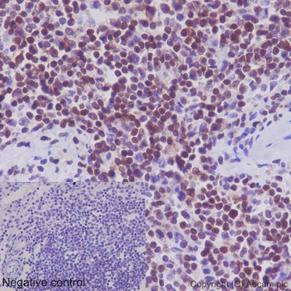
Immunohistochemical analysis of paraffin-embedded Human spleen tissue labeling STAT5b with ab178941 at 1/500 dilution, followed by prediluted HRP Polymer for Rabbit/Mouse IgG. Nucleus staining on lymphocytes of Human spleen is detected. The negative control utilised PBS instead of primary antibody. Counter stained with Hematoxylin.
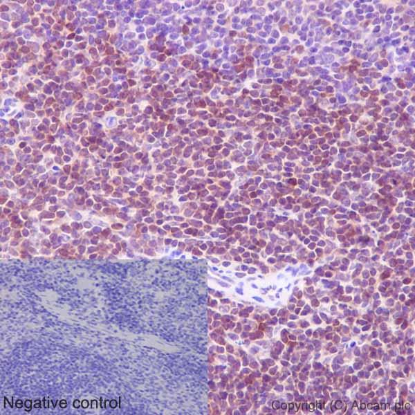
Immunohistochemical analysis of paraffin-embedded Mouse spleen tissue labeling STAT5b with ab178941 at 1/500 dilution, followed by prediluted HRP Polymer for Rabbit/Mouse IgG. Nucleus staining on lymphocytes of Mouse spleen is detected. The negative control utilised PBS instead of primary antibody. Counter stained with Hematoxylin.
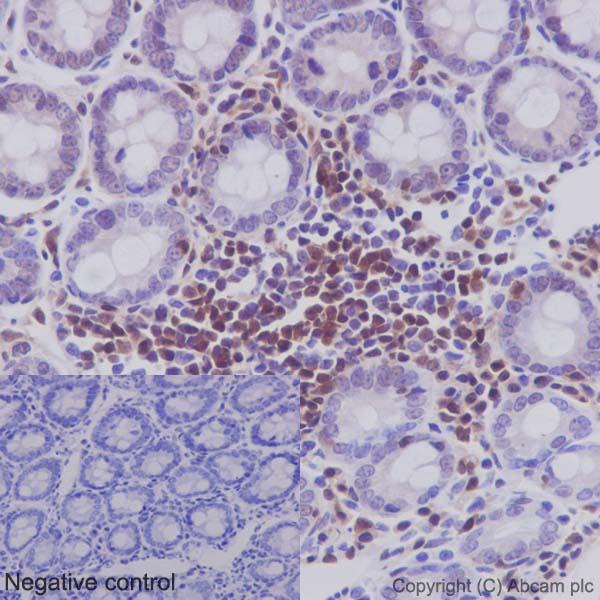
Immunohistochemical analysis of paraffin-embedded Rat colon tissue labeling STAT5b with ab178941 at 1/500 dilution, followed by prediluted HRP Polymer for Rabbit/Mouse IgG. Nucleus staining on lymphocytes and weak nucleus staining on gland epithelium of colon is detected. The negative control utilised PBS insead of primary antibody and the slide is counter stained with Hematoxylin.
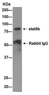
Immunoprecipitation of K562 (Human chronic myelogenous leukemia cells from bone marrow) whole cell extract using ab178941 at 1/40 dilution. Western blot detection of STAT5b utilised ab178941 at 1/2000 dilution and Goat Anti-Rabbit IgG, (H+L), Peroxidase conjugated secondary antibody at 1/1000 dilution. The blocking and dilution buffer was 5% NFDM/TBST.
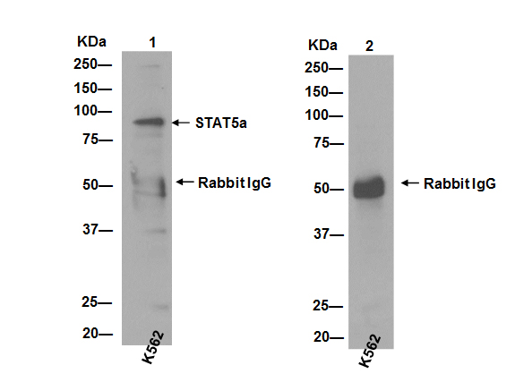
Cross Immunoprecipitation of K562 (Human chronic myelogenous leukemia cells from bone marrow) whole cell extract showing no cross reactivity with STAT5a. Protein captured by anti-STAT5a antibody (ab32042) was detected by the same antibody in WB (image 1) but not by anti-STAT5b, ab178941 (image 2). For WB detection, ab178941 was used at a 1/2000 dilution and Goat Anti-Rabbit IgG, (H+L), Peroxidase conjugated secondary antibody at a 1/1000 dilution. The blocking and dilution buffer was 5% NFDM/TBST.
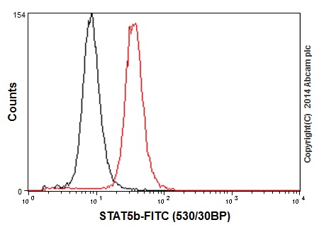
Flow cytometry analysis of 2% paraformaldehyde K562 (Human chronic myelogenous leukemia cells from bone marrow) cells labeling STAT5b with ab178941 at 1:60 dilution (red line). Secondary antibody used is a goat anti rabbit IgG (FITC) at 1:150 dilution. The isotype control is rabbit monoclonal IgG (black line).

![All lanes : Anti-STAT5b [EPR16671] antibody (ab178941) at 1/20000 dilutionLane 1 : K562 (Human chronic myelogenous leukemia cells from bone marrow) whole cell lysatesLane 2 : HeLa (Human epithelial cells from cervix adenocarcinoma) whole cell lysatesLane 3 : Jurkat (Human T cell leukemia cells from peripheral blood) whole cell lysatesLane 4 : Daudi (Human Burkitt's lymphoma cell line) whole cell lysatesLysates/proteins at 20 µg per lane.SecondaryGoat Anti-Rabbit IgG, (H+L), Peroxidase conjugated at 1/1000 dilution](http://www.bioprodhub.com/system/product_images/ab_products/2/sub_5/5875_ab178941-228444-178941.jpg)
![All lanes : Anti-STAT5b [EPR16671] antibody (ab178941) at 1/5000 dilutionLane 1 : Mouse brain lysatesLane 2 : Mouse heart lysatesLane 3 : Mouse kidney lysatesLane 4 : Mouse spleen lysatesLane 5 : Rat brain lysatesLane 6 : Rat heart lysatesLane 7 : Rat kidney lysatesLane 8 : Rat spleen lysatesLane 9 : C6 (Rat glial tumor cells) whole cell lysatesLane 10 : RAW 264.7 (Mouse macrophage cells transformed with Abelson murine leukemia virus) whole cell lysatesLane 11 : PC-12 (Rat adrenal gland pheochromocytoma) whole cell lysatesLane 12 : NIH/3T3 (Mouse embyro fibroblast cells) whole cell lysatesLysates/proteins at 10 µg per lane.SecondaryAnti-Rabbit IgG (HRP), specific to the non-reduced form of IgG at 1/1000 dilution](http://www.bioprodhub.com/system/product_images/ab_products/2/sub_5/5876_ab178941-228447-1789413.jpg)
![All lanes : Anti-STAT5b [EPR16671] antibody (ab178941) at 1/20000 dilutionLane 1 : Human fetal heart lysatesLane 2 : Human fetal kidney lysatesLane 3 : Human fetal spleen lysatesLysates/proteins at 20 µg per lane.SecondaryAnti-Rabbit IgG (HRP), specific to the non-reduced form of IgG at 1/1000 dilution](http://www.bioprodhub.com/system/product_images/ab_products/2/sub_5/5877_ab178941-228445-1789412.jpg)
![Lane 1 : Anti-STAT5b [EPR16671] antibody (ab178941) at 1/1000 dilutionLane 2 : Anti-STAT5a antibody [E289] (ab32043) at 1/5000 dilutionLane 1 : STAT5a recombinant proteinLane 2 : STAT5a recombinant proteindeveloped using the ECL technique](http://www.bioprodhub.com/system/product_images/ab_products/2/sub_5/5878_ab178941-229060-ab178941WB1.jpg)


