![Thymine DNA glycosylase was immunoprecipitated using 0.5mg A431 whole cell extract, 5µg of Rabbit polyclonal to Thymine DNA glycosylase and 50µl of protein G magnetic beads (+). No antibody was added to the control (-).The antibody was incubated under agitation with Protein G beads for 10min, A431 whole cell extract lysate diluted in RIPA buffer was added to each sample and incubated for a further 10min under agitation.Proteins were eluted by addition of 40µl SDS loading buffer and incubated for 10min at 70°C; 10µl of each sample was separated on a SDS PAGE gel, transferred to a nitrocellulose membrane, blocked with 5% BSA and probed with ab106301.Secondary: Mouse monoclonal [SB62a] Secondary Antibody to Rabbit IgG light chain (HRP) (ab99697).Band: 50kDa; Thymine DNA glycosylase](http://www.bioprodhub.com/system/product_images/ab_products/2/sub_5/10848_ab106301-205647-IPVI002ab10630120mMod.jpg)
Thymine DNA glycosylase was immunoprecipitated using 0.5mg A431 whole cell extract, 5µg of Rabbit polyclonal to Thymine DNA glycosylase and 50µl of protein G magnetic beads (+). No antibody was added to the control (-).The antibody was incubated under agitation with Protein G beads for 10min, A431 whole cell extract lysate diluted in RIPA buffer was added to each sample and incubated for a further 10min under agitation.Proteins were eluted by addition of 40µl SDS loading buffer and incubated for 10min at 70°C; 10µl of each sample was separated on a SDS PAGE gel, transferred to a nitrocellulose membrane, blocked with 5% BSA and probed with ab106301.Secondary: Mouse monoclonal [SB62a] Secondary Antibody to Rabbit IgG light chain (HRP) (ab99697).Band: 50kDa; Thymine DNA glycosylase

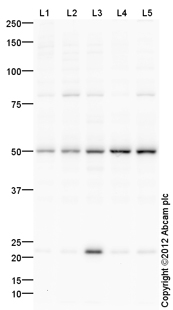
All lanes : Anti-Thymine DNA glycosylase antibody (ab106301) at 1 µg/mlLane 1 : HeLa (Human epithelial carcinoma cell line) Whole Cell LysateLane 2 : Jurkat (Human T cell lymphoblast-like cell line) Whole Cell LysateLane 3 : HEK293 (Human embryonic kidney cell line) Whole Cell LysateLane 4 : A431 (Human epithelial carcinoma cell line) Whole Cell LysateLane 5 : HUES7 (Human embryonic stem cell line) Whole Cell LysateLysates/proteins at 10 µg per lane.SecondaryGoat Anti-Rabbit IgG H&L (HRP) preadsorbed (ab97080) at 1/5000 dilutiondeveloped using the ECL techniquePerformed under reducing conditions.

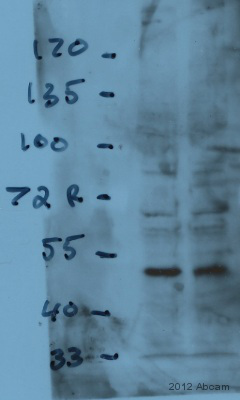
All lanes : Anti-Thymine DNA glycosylase antibody (ab106301) at 1 µg/mlLane 1 : Mouse ES whole cell lysateLane 2 : Mouse ES whole cell lysateLysates/proteins at 100 µg per lane.SecondaryHRP Goat anti-rabbit IgG polyclonal at 1/2000 dilutiondeveloped using the ECL techniquePerformed under reducing conditions.
![Thymine DNA glycosylase was immunoprecipitated using 0.5mg A431 whole cell extract, 5µg of Rabbit polyclonal to Thymine DNA glycosylase and 50µl of protein G magnetic beads (+). No antibody was added to the control (-).The antibody was incubated under agitation with Protein G beads for 10min, A431 whole cell extract lysate diluted in RIPA buffer was added to each sample and incubated for a further 10min under agitation.Proteins were eluted by addition of 40µl SDS loading buffer and incubated for 10min at 70°C; 10µl of each sample was separated on a SDS PAGE gel, transferred to a nitrocellulose membrane, blocked with 5% BSA and probed with ab106301.Secondary: Mouse monoclonal [SB62a] Secondary Antibody to Rabbit IgG light chain (HRP) (ab99697).Band: 50kDa; Thymine DNA glycosylase](http://www.bioprodhub.com/system/product_images/ab_products/2/sub_5/10848_ab106301-205647-IPVI002ab10630120mMod.jpg)

