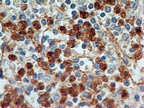
Immunohistochemistry (Formalin/PFA-fixed paraffin-embedded sections) analysis of Human spleen tissue labelling TLR9 with ab134368 at 5µg/ml. Staining was enhanced by boiling tissue sections in 10mM sodium citrate buffer, pH6.0 for 10-20 minutes followed by cooling at room temperature for 20 minutes.
![All lanes : Anti-TLR9 antibody [26C593.2] (ab134368) at 3 µg/mlLane 1 : Human Peripheral Blood Mononuclear Cells (PBMCs) Lane 2 : Human intestine tissue lysateLane 3 : Mouse intestine tissue lysateLane 4 : Rat intestine tissue lysateLysates/proteins at 40 µg per lane.SecondaryGoat anti-mouse HRP conjugate](http://www.bioprodhub.com/system/product_images/ab_products/2/sub_5/11794_TLR9-Primary-antibodies-ab134368-1.jpg)
All lanes : Anti-TLR9 antibody [26C593.2] (ab134368) at 3 µg/mlLane 1 : Human Peripheral Blood Mononuclear Cells (PBMCs) Lane 2 : Human intestine tissue lysateLane 3 : Mouse intestine tissue lysateLane 4 : Rat intestine tissue lysateLysates/proteins at 40 µg per lane.SecondaryGoat anti-mouse HRP conjugate

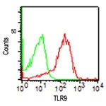
Flow Cytometric analysis of TLR9 in Human PBMC cells labelled with ab134368 at 0.5µg (Red), or isotype control antibody (green).

Flow Cytometric analysis of TLR9 in Ramos cells labelled with ab134368 at 0.1 µg (Red), no antibody (blue) or isotype control antibody (green). Goat anti-mouse IgG FITC secondary was used.

Immunohistochemical analysis of TLR9 in frozen sections of Human lung adenocarcinoma tissue, using ab134368 at a 1/100 dilution.

Immunohistochemical analysis of TLR9 in frozen sections of A549 cells, using ab134368 at a 1/100 dilution.

Immunohistochemical analysis of TLR9 in frozen sections of malignant Human lung tissue, using ab134368 at a 1/100 dilution.

![All lanes : Anti-TLR9 antibody [26C593.2] (ab134368) at 3 µg/mlLane 1 : Human Peripheral Blood Mononuclear Cells (PBMCs) Lane 2 : Human intestine tissue lysateLane 3 : Mouse intestine tissue lysateLane 4 : Rat intestine tissue lysateLysates/proteins at 40 µg per lane.SecondaryGoat anti-mouse HRP conjugate](http://www.bioprodhub.com/system/product_images/ab_products/2/sub_5/11794_TLR9-Primary-antibodies-ab134368-1.jpg)


