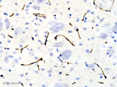
Anti-Transferrin Receptor antibody
| Name | Anti-Transferrin Receptor antibody |
|---|---|
| Supplier | Abcam |
| Catalog | ab84036 |
| Prices | $398.00 |
| Sizes | 100 µg |
| Host | Rabbit |
| Clonality | Polyclonal |
| Isotype | IgG |
| Applications | WB ICC/IF ICC/IF IHC-F IHC-P |
| Species Reactivities | Mouse, Human, Dog, Pig, Orangutan |
| Antigen | Synthetic peptide conjugated to KLH derived from within residues 1 - 100 of Human Transferrin Receptor |
| Description | Rabbit Polyclonal |
| Gene | TFRC |
| Conjugate | Unconjugated |
| Supplier Page | Shop |
Product images
Product References
Transferrin receptor specific nanocarriers conjugated with functional 7peptide - Transferrin receptor specific nanocarriers conjugated with functional 7peptide
Du W, Fan Y, Zheng N, He B, Yuan L, Zhang H, Wang X, Wang J, Zhang X, Zhang Q. Biomaterials. 2013 Jan;34(3):794-806.
The memory-enhancing effects of hippocampal estrogen receptor activation involve - The memory-enhancing effects of hippocampal estrogen receptor activation involve
Boulware MI, Heisler JD, Frick KM. J Neurosci. 2013 Sep 18;33(38):15184-94.
Interaction of ganglioside GD3 with an EGF receptor sustains the self-renewal - Interaction of ganglioside GD3 with an EGF receptor sustains the self-renewal
Wang J, Yu RK. Proc Natl Acad Sci U S A. 2013 Nov 19;110(47):19137-42. doi:
Terminal differentiation and loss of tumorigenicity of human cancers via - Terminal differentiation and loss of tumorigenicity of human cancers via
Zhang X, Cruz FD, Terry M, Remotti F, Matushansky I. Oncogene. 2013 May 2;32(18):2249-60, 2260.e1-21.


