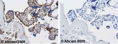
Ab44928 staining human normal placenta. Staining is localized to the cytoplamsmLeft panel: with primary antibody at 2 ug/ml. Right panel: isotype control.Sections were stained using an automated system DAKO Autostainer Plus , at room temperature. Sections were rehydrated and antigen retrieved with the Dako 3-in-1 AR buffers EDTA pH 9.0 in a DAKO PT Link. Slides were peroxidase blocked in 3% H2O2 in methanol for 10 minutes. They were then blocked with Dako Protein block for 10 minutes (containing casein 0.25% in PBS) then incubated with primary antibody for 20 minutes and detected with Dako Envision Flex amplification kit for 30 minutes. Colorimetric detection was completed with Diaminobenzidine for 5 minutes. Slides were counterstained with Haematoxylin and coverslipped under DePeX. Please note that for manual staining we recommend to optimize the primary antibody concentration and incubation time (overnight incubation), and amplification may be required.

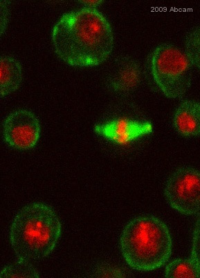
ab44928 staining Tubulin in Drosophila S2 cells by ICC/IF (Immunocytochemistry/immunofluorescence). Cells were fixed with methanol and blocked with 5% BSA for 15 minutes at Room temperature. Samples were incubated with primary antibody (1/2000 in 5% BSA + PBST) for 2 hours. Ab6785 (1/1000) was used as secondary antibody.See Abreview
![All lanes : Anti-Tubulin antibody [DM1A +DM1B] (ab44928) at 1/5000 dilutionLane 1 : Whole cell lysate prepared from human colon cellsLane 2 : Whole cell lysate prepared from human colon cellsLysates/proteins at 20 µg per lane.SecondaryHRP sheep anti-mouse polyclonal at 1/10000 dilutiondeveloped using the ECL technique](http://www.bioprodhub.com/system/product_images/ab_products/2/sub_5/16332_Tubulin-Primary-antibodies-ab44928-7.jpg)
All lanes : Anti-Tubulin antibody [DM1A +DM1B] (ab44928) at 1/5000 dilutionLane 1 : Whole cell lysate prepared from human colon cellsLane 2 : Whole cell lysate prepared from human colon cellsLysates/proteins at 20 µg per lane.SecondaryHRP sheep anti-mouse polyclonal at 1/10000 dilutiondeveloped using the ECL technique
![All lanes : Anti-Tubulin antibody [DM1A +DM1B] (ab44928) at 5 µg/mlLane 1 : HeLa (Human epithelial carcinoma cell line) Whole Cell LysateLane 2 : NIH 3T3 (Mouse embryonic fibroblast cell line) Whole Cell Lysate Lane 3 : PC12 (Rat adrenal pheochromocytoma cell line) Whole Cell Lysate Lysates/proteins at 20 µg per lane.SecondaryGoat Anti-Mouse IgG H&L (HRP) preadsorbed (ab97040) at 1/5000 dilutiondeveloped using the ECL techniquePerformed under reducing conditions.](http://www.bioprodhub.com/system/product_images/ab_products/2/sub_5/16333_Tubulin-Primary-antibodies-ab44928-9.jpg)
All lanes : Anti-Tubulin antibody [DM1A +DM1B] (ab44928) at 5 µg/mlLane 1 : HeLa (Human epithelial carcinoma cell line) Whole Cell LysateLane 2 : NIH 3T3 (Mouse embryonic fibroblast cell line) Whole Cell Lysate Lane 3 : PC12 (Rat adrenal pheochromocytoma cell line) Whole Cell Lysate Lysates/proteins at 20 µg per lane.SecondaryGoat Anti-Mouse IgG H&L (HRP) preadsorbed (ab97040) at 1/5000 dilutiondeveloped using the ECL techniquePerformed under reducing conditions.
![Overlay histogram showing HeLa cells stained with ab44928 (red line). The cells were fixed with 80% methanol (5 min) and then permeabilized with 0.1% PBS-Tween for 20 min. The cells were then incubated in 1x PBS / 10% normal goat serum / 0.3M glycine to block non-specific protein-protein interactions. The cells were then incubated with the antibody (ab449287, 1µg/1x106 cells) for 30 min at 22ºC. The secondary antibody used was DyLight® 488 goat anti-mouse IgG (H+L) (ab96879) at 1/500 dilution for 30 min at 22ºC. Isotype control antibody (black line) was mouse IgG1 [ICIGG1] (ab91353, 2µg/1x106 cells ) used under the same conditions. Acquisition of >5,000 events was performed. This antibody gave a positive signal in HeLa cells fixed with 4% paraformaldehyde (10 min)/permeabilized in 0.1% PBS-Tween used under the same conditions.](http://www.bioprodhub.com/system/product_images/ab_products/2/sub_5/16334_Tubulin-Primary-antibodies-ab44928-10.jpg)
Overlay histogram showing HeLa cells stained with ab44928 (red line). The cells were fixed with 80% methanol (5 min) and then permeabilized with 0.1% PBS-Tween for 20 min. The cells were then incubated in 1x PBS / 10% normal goat serum / 0.3M glycine to block non-specific protein-protein interactions. The cells were then incubated with the antibody (ab449287, 1µg/1x106 cells) for 30 min at 22ºC. The secondary antibody used was DyLight® 488 goat anti-mouse IgG (H+L) (ab96879) at 1/500 dilution for 30 min at 22ºC. Isotype control antibody (black line) was mouse IgG1 [ICIGG1] (ab91353, 2µg/1x106 cells ) used under the same conditions. Acquisition of >5,000 events was performed. This antibody gave a positive signal in HeLa cells fixed with 4% paraformaldehyde (10 min)/permeabilized in 0.1% PBS-Tween used under the same conditions.


![All lanes : Anti-Tubulin antibody [DM1A +DM1B] (ab44928) at 1/5000 dilutionLane 1 : Whole cell lysate prepared from human colon cellsLane 2 : Whole cell lysate prepared from human colon cellsLysates/proteins at 20 µg per lane.SecondaryHRP sheep anti-mouse polyclonal at 1/10000 dilutiondeveloped using the ECL technique](http://www.bioprodhub.com/system/product_images/ab_products/2/sub_5/16332_Tubulin-Primary-antibodies-ab44928-7.jpg)
![All lanes : Anti-Tubulin antibody [DM1A +DM1B] (ab44928) at 5 µg/mlLane 1 : HeLa (Human epithelial carcinoma cell line) Whole Cell LysateLane 2 : NIH 3T3 (Mouse embryonic fibroblast cell line) Whole Cell Lysate Lane 3 : PC12 (Rat adrenal pheochromocytoma cell line) Whole Cell Lysate Lysates/proteins at 20 µg per lane.SecondaryGoat Anti-Mouse IgG H&L (HRP) preadsorbed (ab97040) at 1/5000 dilutiondeveloped using the ECL techniquePerformed under reducing conditions.](http://www.bioprodhub.com/system/product_images/ab_products/2/sub_5/16333_Tubulin-Primary-antibodies-ab44928-9.jpg)
![Overlay histogram showing HeLa cells stained with ab44928 (red line). The cells were fixed with 80% methanol (5 min) and then permeabilized with 0.1% PBS-Tween for 20 min. The cells were then incubated in 1x PBS / 10% normal goat serum / 0.3M glycine to block non-specific protein-protein interactions. The cells were then incubated with the antibody (ab449287, 1µg/1x106 cells) for 30 min at 22ºC. The secondary antibody used was DyLight® 488 goat anti-mouse IgG (H+L) (ab96879) at 1/500 dilution for 30 min at 22ºC. Isotype control antibody (black line) was mouse IgG1 [ICIGG1] (ab91353, 2µg/1x106 cells ) used under the same conditions. Acquisition of >5,000 events was performed. This antibody gave a positive signal in HeLa cells fixed with 4% paraformaldehyde (10 min)/permeabilized in 0.1% PBS-Tween used under the same conditions.](http://www.bioprodhub.com/system/product_images/ab_products/2/sub_5/16334_Tubulin-Primary-antibodies-ab44928-10.jpg)