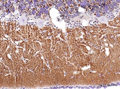
IHC-P image of VGlut1 staining on goat cerebellum section using ab104898 (1:4000). The sections were deparaffinized and subjected to heat mediated antigen retrieval using citric acid. The sections were blocked using 1% BSA for 10 mins at 21°C. ab104898 was diluted 1:4000 and incubated with the sections for 2 hours at 21°C. The secondary antibody used was goat polyclonal to rabbit IgG conjugated to biotin (undiluted).See Abreview
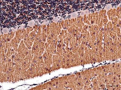
IHC-P image of VGlut1 staining on rat cerebellum section using ab104898 (1:4000). The sections were deparaffinized and subjected to heat mediated antigen retrieval using citric acid. The sections were blocked using 1% BSA for 10 mins at 21°C. ab104898 was diluted 1:4000 and incubated with the sections for 2 hours at 21°C. The secondary antibody used was goat polyclonal to rabbit IgG conjugated to biotin (undiluted).
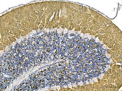
IHC-P image of VGlut1 staining on mouse cerebellum section using ab104898 (1:4000). The sections were deparaffinized and subjected to heat mediated antigen retrieval using citric acid. The sections were blocked using 1% BSA for 10 mins at 21°C. ab104898 was diluted 1:4000 and incubated with the sections for 2 hours at 21°C. The secondary antibody used was goat polyclonal to rabbit IgG conjugated to biotin (1:250).
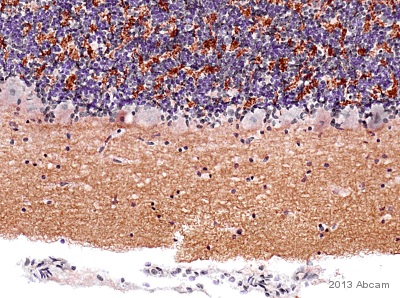
IHC-P image of VGlut1 staining on Cat cerebellum section using ab104898 (1:4000). The sections were deparaffinized and subjected to heat mediated antigen retrieval using citric acid. The sections were blocked using 1% BSA for 10 mins at 21°C. ab104898 was diluted 1:4000 and incubated with the sections for 2 hours at 21°C. The secondary antibody used was goat polyclonal to rabbit IgG conjugated to biotin (1:250).
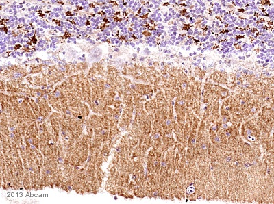
IHC-P image of VGlut1 staining on dog cerebellum section using ab104898 (1:4000). The sections were deparaffinized and subjected to heat mediated antigen retrieval using citric acid. The sections were blocked using 1% BSA for 10 mins at 21°C. ab104898 was diluted 1:4000 and incubated with the sections for 2 hours at 21°C. The secondary antibody used was goat polyclonal to rabbit IgG conjugated to biotin (1:250).
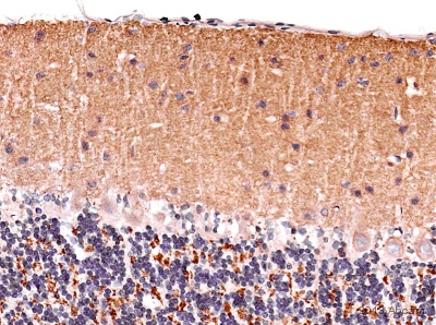
IHC-P image of VGlut1 staining on marmoset cerebellum section using ab104898 (1:4000). The sections were deparaffinized and subjected to heat mediated antigen retrieval using citric acid. The sections were blocked using 1% BSA for 10 mins at 21°C. ab104898 was diluted 1:4000 and incubated with the sections for 2 hours at 21°C. The secondary antibody used was goat polyclonal to rabbit IgG conjugated to biotin (1:250).


