Anti-Vimentin antibody [V9]
| Name | Anti-Vimentin antibody [V9] |
|---|---|
| Supplier | Abcam |
| Catalog | ab80667 |
| Prices | $385.00 |
| Sizes | 100 µl |
| Host | Mouse |
| Clonality | Monoclonal |
| Isotype | IgG1 |
| Clone | V9 |
| Applications | ICC/IF ICC/IF IHC-P FC |
| Species Reactivities | Mouse, Rat, Rabbit, Horse, Chicken, Hamster, Bovine, Cat, Dog, Human, Pig, Monkey, Rat |
| Antigen | Purified Vimentin from pig eye lens |
| Description | Mouse Monoclonal |
| Gene | VIM |
| Conjugate | Unconjugated |
| Supplier Page | Shop |
Product images
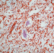 ab80667 at 1/100 dilution staining Vimentin in osteosarcoma by Immunohistochemistry, Formalin fixed, Paraffin-embedded tissue using peroxidase-conjugate and AEC chromogen. Note cytoplasmic staining of tumor cells.
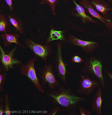 ICC/IF image of ab80667 stained HeLa cells. The cells were 4% paraformaldehyde fixed (10 min) and then incubated in 1%BSA / 10% normal goat serum / 0.3M glycine in 0.1% PBS-Tween for 1h to permeabilise the cells and block non-specific protein-protein interactions. The cells were then incubated with the antibody (ab80667, 1µg/ml) overnight at +4°C. The secondary antibody (green) was ab96879, DyLight® 488 goat anti-mouse IgG (H+L) used at a 1/250 dilution for 1h. Alexa Fluor® 594 WGA was used to label plasma membranes (red) at a 1/200 dilution for 1h. DAPI was used to stain the cell nuclei (blue) at a concentration of 1.43µM.
![Overlay histogram showing HeLa cells stained with ab80667 (red line). The cells were fixed with 80% methanol (5 min) and then permeabilized with 0.1% PBS-Tween for 20 min. The cells were then incubated in 1x PBS / 10% normal goat serum / 0.3M glycine to block non-specific protein-protein interactions followed by the antibody (ab80667, 0.1μg/1x106 cells) for 30 min at 22°C. The secondary antibody used was Alexa Fluor® 488 goat anti-mouse IgG (H&L) (ab150113) at 1/2000 dilution for 30 min at 22°C. Isotype control antibody (black line) was mouse IgG1 [ICIGG1] (ab91353, 1μg/1x106 cells) used under the same conditions. Unlabelled sample (blue line) was also used as a control. Acquisition of >5,000 events were collected using a 20mW Argon ion laser (488nm) and 525/30 bandpass filter.](http://www.bioprodhub.com/system/product_images/ab_products/2/sub_5/19949_ab80667-3-ab80667FC.jpg) Overlay histogram showing HeLa cells stained with ab80667 (red line). The cells were fixed with 80% methanol (5 min) and then permeabilized with 0.1% PBS-Tween for 20 min. The cells were then incubated in 1x PBS / 10% normal goat serum / 0.3M glycine to block non-specific protein-protein interactions followed by the antibody (ab80667, 0.1μg/1x106 cells) for 30 min at 22°C. The secondary antibody used was Alexa Fluor® 488 goat anti-mouse IgG (H&L) (ab150113) at 1/2000 dilution for 30 min at 22°C. Isotype control antibody (black line) was mouse IgG1 [ICIGG1] (ab91353, 1μg/1x106 cells) used under the same conditions. Unlabelled sample (blue line) was also used as a control. Acquisition of >5,000 events were collected using a 20mW Argon ion laser (488nm) and 525/30 bandpass filter.
Overlay histogram showing HeLa cells stained with ab80667 (red line). The cells were fixed with 80% methanol (5 min) and then permeabilized with 0.1% PBS-Tween for 20 min. The cells were then incubated in 1x PBS / 10% normal goat serum / 0.3M glycine to block non-specific protein-protein interactions followed by the antibody (ab80667, 0.1μg/1x106 cells) for 30 min at 22°C. The secondary antibody used was Alexa Fluor® 488 goat anti-mouse IgG (H&L) (ab150113) at 1/2000 dilution for 30 min at 22°C. Isotype control antibody (black line) was mouse IgG1 [ICIGG1] (ab91353, 1μg/1x106 cells) used under the same conditions. Unlabelled sample (blue line) was also used as a control. Acquisition of >5,000 events were collected using a 20mW Argon ion laser (488nm) and 525/30 bandpass filter.
Product References
Development of cytoskeleton in neuroectodermally derived epithelial and muscle - Development of cytoskeleton in neuroectodermally derived epithelial and muscle
Uusitalo M, Kivela T. Invest Ophthalmol Vis Sci. 1995 Dec;36(13):2584-91.
Molecular genetic analysis of flow-sorted ovarian tumour cells: improved - Molecular genetic analysis of flow-sorted ovarian tumour cells: improved
Abeln EC, Corver WE, Kuipers-Dijkshoorn NJ, Fleuren GJ, Cornelisse CJ. Br J Cancer. 1994 Aug;70(2):255-62.