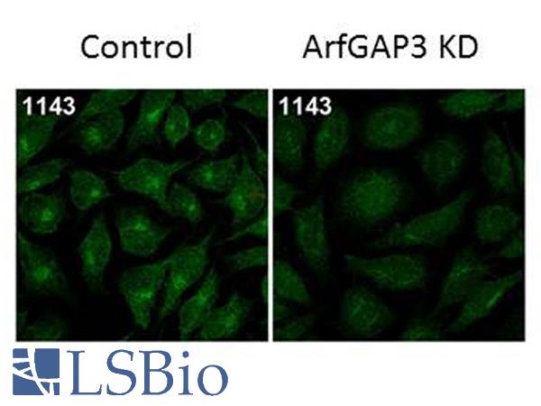
Immunofluorescence Microscopy of Rabbit Anti-ArfGAP3 Antibody. Tissue: HeLa Whole Cell. Fixation: MeOH. Antigen retrieval: not required. Primary antibody: ArfGAP3 antibody at 1:100 for 1 h at RT. Secondary antibody: Fluorescein rabbit secondary antibody at 1:10,000 for 45 min at RT. Localization: ArfGAP3 is cytoplasmic. Staining: ArfGAP3 as green fluorescent signal.

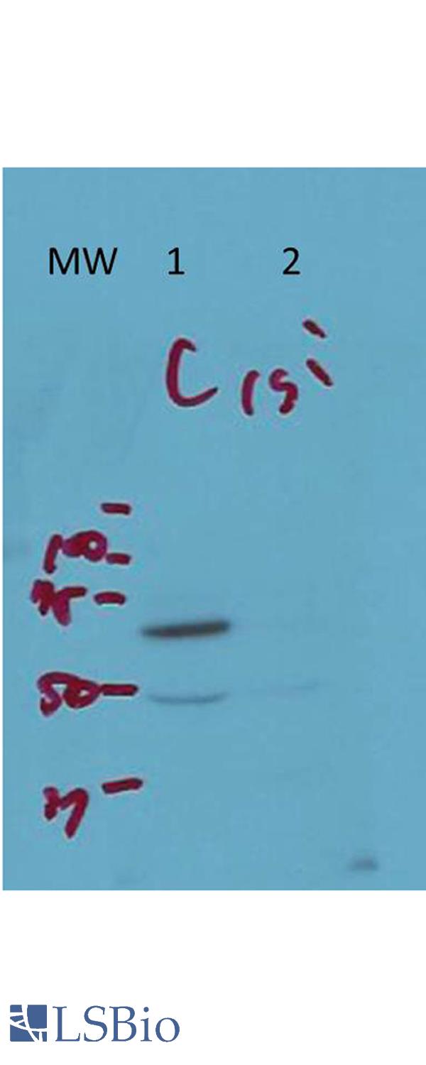
Western Blot of Rabbit Anti-ArfGAP3 Antibody. Lane 1 (C): HeLa Whole Cell. Lane 2 (si): HeLa Whole Cell siRNA treated. Load: 10 ug per lane. Primary antibody: ArfGAP3 antibody at 1:1000 for overnight at 4 degrees C. Secondary antibody: IRDye800 alpha rabbit secondary antibody at 1:10,000 for 45 min at RT. Block: 5% BLOTTO overnight at 4 degrees C. Predicted/Observed size: 57 kDa for endogenous Arf-GAP3. Other band(s): non-specific band ~50kDa.

