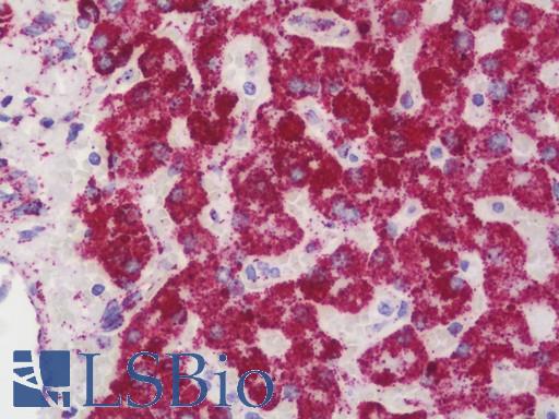
Anti-ATP5B / ATP Synthase Beta antibody IHC staining of human liver. Immunohistochemistry of formalin-fixed, paraffin-embedded tissue after heat-induced antigen retrieval.

Anti-ATP5B / ATP Synthase Beta antibody IHC staining of human kidney. Immunohistochemistry of formalin-fixed, paraffin-embedded tissue after heat-induced antigen retrieval. Antibody LS-B10700 dilution 1:75.
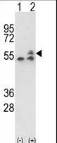
Western blot of ATP5B (arrow) using rabbit polyclonal ATP5B Antibody. 293 cell lysates (2 ug/lane) either nontransfected (Lane 1) or transiently transfected with the ATP5B gene (Lane 2).

Western blot of ATP5B Antibody antibody pre-incubated without(lane 1) and with(lane 2) blocking peptide in WiDr cell line lysate. ATP5B (arrow) was detected using the purified antibody.

ATP5B Antibody flow cytometry of WiDr cells (right histogram) compared to a negative control cell (left histogram). FITC-conjugated goat-anti-rabbit secondary antibodies were used for the analysis.