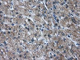
![IHC of paraffin-embedded liver tissue using anti-ATP5B mouse monoclonal antibody. (Dilution 1:50).](http://www.bioprodhub.com/system/product_images/ab_products/3/sub_1/10979_37612_79139.jpg)
IHC of paraffin-embedded liver tissue using anti-ATP5B mouse monoclonal antibody. (Dilution 1:50).
![IHC of paraffin-embedded Kidney tissue using anti-ATP5B mouse monoclonal antibody. (Dilution 1:50).](http://www.bioprodhub.com/system/product_images/ab_products/3/sub_1/10980_37611_79138.jpg)
IHC of paraffin-embedded Kidney tissue using anti-ATP5B mouse monoclonal antibody. (Dilution 1:50).
![IHC of paraffin-embedded Carcinoma of liver tissue using anti-ATP5B mouse monoclonal antibody. (Dilution 1:50).](http://www.bioprodhub.com/system/product_images/ab_products/3/sub_1/10981_37613_79140.jpg)
IHC of paraffin-embedded Carcinoma of liver tissue using anti-ATP5B mouse monoclonal antibody. (Dilution 1:50).
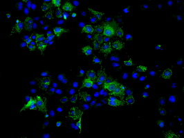
![Anti-ATP5B mouse monoclonal antibody immunofluorescent staining of COS7 cells transiently transfected by pCMV6-ENTRY ATP5B.](http://www.bioprodhub.com/system/product_images/ab_products/3/sub_1/10982_37614_79141.jpg)
Anti-ATP5B mouse monoclonal antibody immunofluorescent staining of COS7 cells transiently transfected by pCMV6-ENTRY ATP5B.
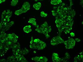
![Immunofluorescent staining of HepG2 cells using anti-ATP5B mouse monoclonal antibody.](http://www.bioprodhub.com/system/product_images/ab_products/3/sub_1/10983_37615_79142.jpg)
Immunofluorescent staining of HepG2 cells using anti-ATP5B mouse monoclonal antibody.
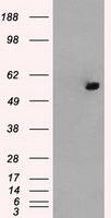
![HEK293T cells were transfected with the pCMV6-ENTRY control (Left lane) or pCMV6-ENTRY ATP5B (Right lane) cDNA for 48 hrs and lysed. Equivalent amounts of cell lysates (5 ug per lane) were separated by SDS-PAGE and immunoblotted with anti-ATP5B.](http://www.bioprodhub.com/system/product_images/ab_products/3/sub_1/10984_37610_79137.jpg)
HEK293T cells were transfected with the pCMV6-ENTRY control (Left lane) or pCMV6-ENTRY ATP5B (Right lane) cDNA for 48 hrs and lysed. Equivalent amounts of cell lysates (5 ug per lane) were separated by SDS-PAGE and immunoblotted with anti-ATP5B.
![Flow cytometry of Jurkat cells, using anti-ATP5B antibody, (Red) compared to a nonspecific negative control antibody (Blue).](http://www.bioprodhub.com/system/product_images/ab_products/3/sub_1/10985_37617_79144.jpg)
Flow cytometry of Jurkat cells, using anti-ATP5B antibody, (Red) compared to a nonspecific negative control antibody (Blue).
![Flow cytometry of HeLa cells, using anti-ATP5B antibody, (Red) compared to a nonspecific negative control antibody (Blue).](http://www.bioprodhub.com/system/product_images/ab_products/3/sub_1/10986_37616_79143.jpg)
Flow cytometry of HeLa cells, using anti-ATP5B antibody, (Red) compared to a nonspecific negative control antibody (Blue).