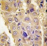
Formalin-fixed and paraffin-embedded human hepatocarcinoma tissue reacted with Autophagy APG7L antibody , which was peroxidase-conjugated to the secondary antibody, followed by DAB staining. This data demonstrates the use of this antibody for immunohistochemistry; clinical relevance has not been evaluated.
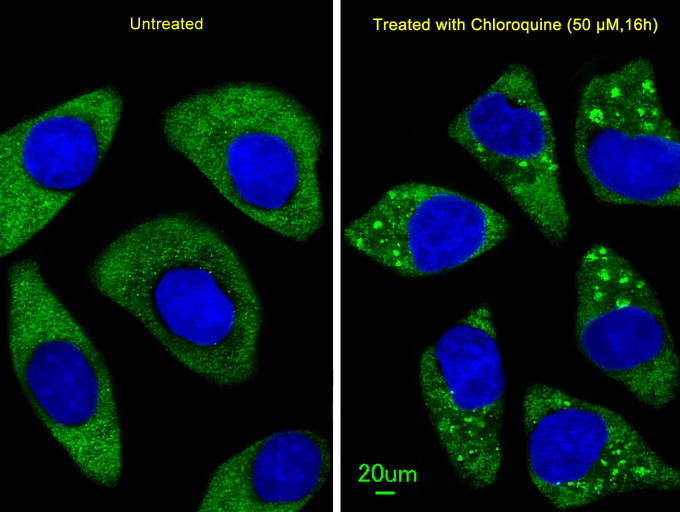
Immunofluorescent of U251 cells, using ATG7 Antibody. U251 cells(right) were treated with Chloroquine (50 mu M,16h). Antibody was diluted at 1:25 dilution. Alexa Fluor 488-conjugated goat anti-rabbit lgG at 1:400 dilution was used as the secondary antibody (green). DAPI was used to stain the cell nuclear (blue).
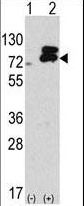
Western blot of APG7L antibody in 293 cell line lysates transiently transfected with the ATG7 gene(2 ug/lane).
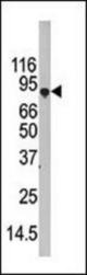
Western blot of APG7L antibody in 293 cell line lysate (35 ug/lane). APG7L (arrow) was detected using the purified antibody.