Anti-HECW2 Antibody
| Name | Anti-HECW2 Antibody |
|---|---|
| Supplier | Cohesion Biosciences |
| Catalog | CPA3425 |
| Prices | $110.00, $220.00, $350.00 |
| Sizes | 30 µl, 100 µl, 200 µl |
| Host | Rabbit |
| Clonality | Polyclonal |
| Applications | WB ICC/IF |
| Species Reactivities | Human, Mouse |
| Antigen | KLH-conjugated synthetic peptide encompassing a sequence within the center region of human HECW2. The exact sequence is proprietary. |
| Purity/Format | The antibody was purified by immunogen affinity chromatography. |
| Blocking Peptide | HECW2 Blocking Peptide |
| Description | Rabbit Polyclonal |
| Gene | HECW2 |
| Supplier Page | Shop |
Product images
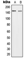 Western blot analysis of HECW2 expression in A549 (A); NIH3T3 (B) whole cell lysates.
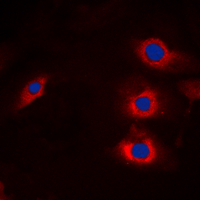 Immunofluorescent analysis of HECW2 staining in A549 cells. Formalin-fixed cells were permeabilized with 0.1% Triton X-100 in TBS for 5-10 minutes and blocked with 3% BSA-PBS for 30 minutes at room temperature. Cells were probed with the primary antibody in 3% BSA-PBS and incubated overnight at 4 C in a humidified chamber. Cells were washed with PBST and incubated with a DyLight 594-conjugated secondary antibody (red) in PBS at room temperature in the dark. DAPI was used to stain the cell nuclei (blue).