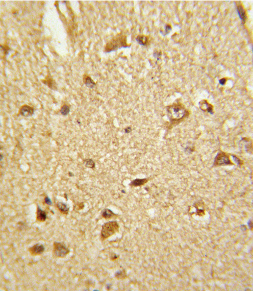
Formalin-fixed and paraffin-embedded human brain tissue reacted with CBS Antibody , which was peroxidase-conjugated to the secondary antibody, followed by DAB staining. This data demonstrates the use of this antibody for immunohistochemistry; clinical relevance has not been evaluated.
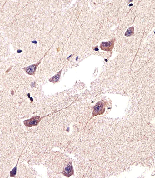
Immunohistochemical of paraffin-embedded H. brain section using CBS Antibody. Antibody was diluted at 1:25 dilution. A peroxidase-conjugated goat anti-rabbit IgG at 1:400 dilution was used as the secondary antibody, followed by DAB staining.

Confocal immunofluorescent of CBS Antibody with 293 cell followed by Alexa Fluor 488-conjugated goat anti-rabbit lgG (green). Actin filaments have been labeled with Alexa Fluor 555 phalloidin (red). DAPI was used to stain the cell nuclear (blue).

CBS Antibody western blot of Raji cell line, rat brain and cerebellum tissue lysates (35 ug/lane). The CBS antibody detected the CBS protein (arrow).
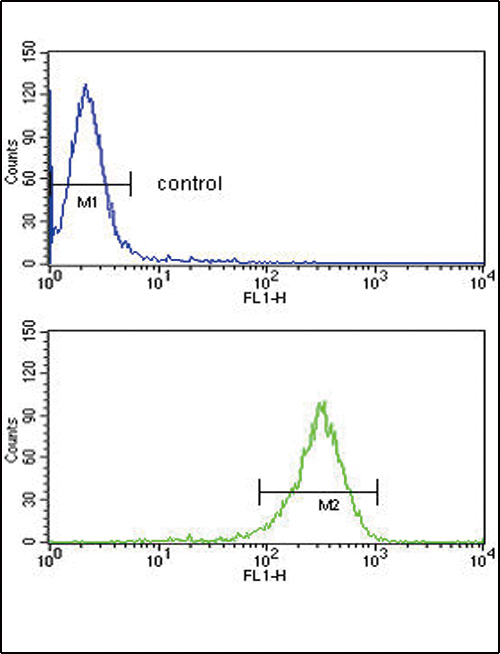
CBS Antibody flow cytometry of 293 cells (bottom histogram) compared to a negative control cell (top histogram). FITC-conjugated goat-anti-rabbit secondary antibodies were used for the analysis.