FoxO3a (75D8) Rabbit mAb
| Name | FoxO3a (75D8) Rabbit mAb |
|---|---|
| Supplier | Cell Signaling Technology |
| Catalog | 2497 |
| Prices | $246.00 |
| Sizes | 100 µl (10 western blots) |
| Host | Rabbit |
| Clonality | Monoclonal |
| Isotype | IgG |
| Clone | 75D8 |
| Applications | WB IP ICC/IF |
| Species Reactivities | Human, Mouse, Rat, Monkey |
| Antigen | Monoclonal antibody is produced by immunizing animals with a synthetic peptide corresponding to residues surrounding Glu50 of human FoxO3a. |
| Description | Rabbit Monoclonal |
| Gene | FOXO3 |
| Supplier Page | Shop |
Product images
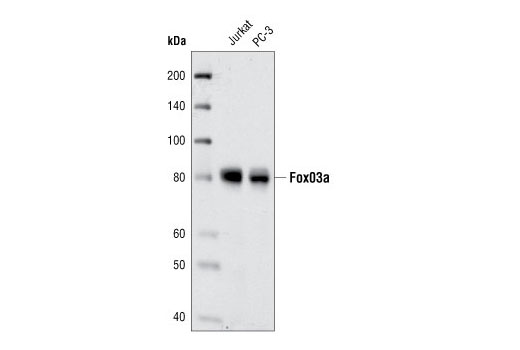 Western blot analysis of extracts from Jurkat and PC3 cells using FoxO3a (75D8) Rabbit mAb.
 Western blot analysis of extracts from wild type (+/+) and FoxO3 knock-out (-/-) mouse brain, using FoxO3a (75D8) Rabbit mAb (upper) and FoxO1 (C29H4) Rabbit mAb #2880 (lower). (FoxO3 knock-out (-/-) mouse brain was kindly provided by Dr. Ron DePinho, Dana-Farber Cancer Institute, Boston, MA).
 Confocal immunofluorescent analysis of SH-SY5Y cells, IGF-I treated (left) or LY294002-treated, using FoxO3a (75D8) Rabbit mAb (green). Actin filaments have been labeled with Alexa Fluor® 555 phalloidin (red). Blue pseudocolor = DRAQ5® #4084 (fluorescent DNA dye).