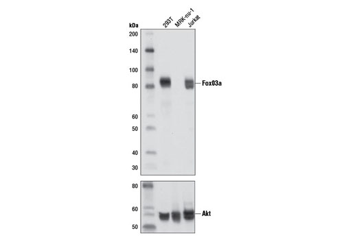
Western blot analysis of extracts from 293T, MRK-nu-1 and Jurkat cells using FoxO3a (D19A7) Rabbit mAb (upper) and Akt (pan) (C67E7) Rabbit mAb #4691 (lower).

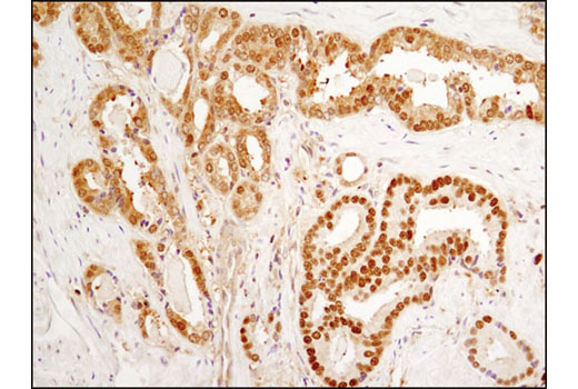
Immunohistochemical analysis of paraffin-embedded human breast carcinoma using FoxO3a (D19A7) Rabbit mAb.

Immunohistochemical analysis of paraffin-embedded PC-3 (upper) and MRK-nu-1 (lower) cell pellets, treated with Human Insulin-like Growth Factor I (hIGF-I) #8917 (left) or LY294002 #9901 (right), using FoxO3a (D19A7) Rabbit mAb.

Immunohistochemical analysis of paraffin-embedded human prostate carcinoma using FoxO3a (D19A7) Rabbit mAb.

Immunohistochemical analysis of paraffin-embedded metastatic SKOV3 tumor in mouse lung using FoxO3a (D19A7) Rabbit mAb. Note nuclear staining in adjacent normal lung.

Confocal immunofluorescent analysis of PC-3 cells, treated with Human Insulin-like Growth Factor I (hIGF-I) #8917 (left) or LY294002 #9901 (right), using FoxO3a (D19A7) Rabbit mAb (green). Actin filaments were labeled with DY-554 phalloidin (red).

Flow cytometric analysis of MCF7 cells (green) and MRK-NU-1 cells (blue) using FoxO3a (D19A7) Rabbit mAb. Anti-rabbit (H+L), F(ab')2 Fragment (PE Conjugate) #8885 was used as a secondary antibody.


