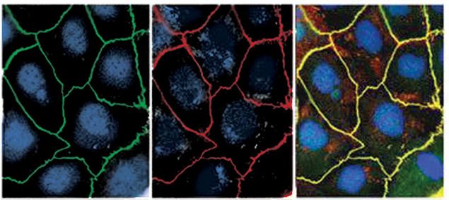
Immunofluorescent staining of Caco-2 cells using Rb anti-ZO-1 (Mid) (Cat. no. 40-2200). Image courtesy of Jacey Bennis and Dr. James Anderson, University of North Carolina at Chapel Hill, NC.
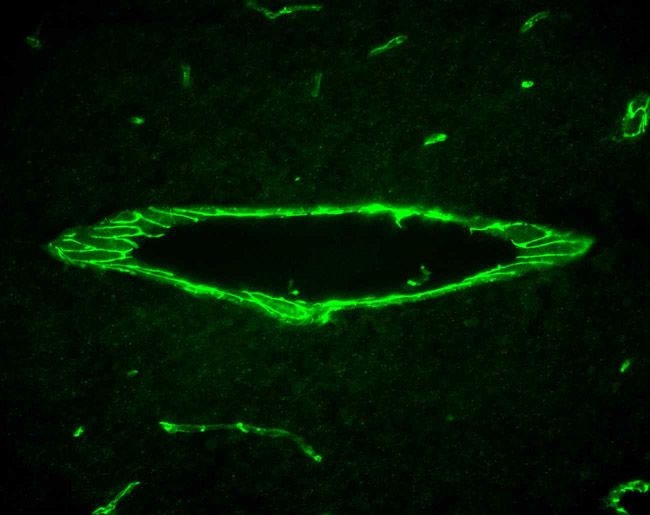
Immunofluorescent staining of blood vessels in mouse heart tissue using Rb anti-ZO-1 (Mid) (Cat. No. 40-2200). Image courtesy of James I. Nagy, PhD, University of Manitoba, Canada.
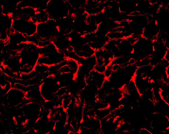
Immunofluorescent staining of mouse liver tissue using Rb anti-ZO-1 (Mid) (Cat. No. 40-2200). Image courtesy of James I. Nagy, PhD, University of Manitoba, Canada.
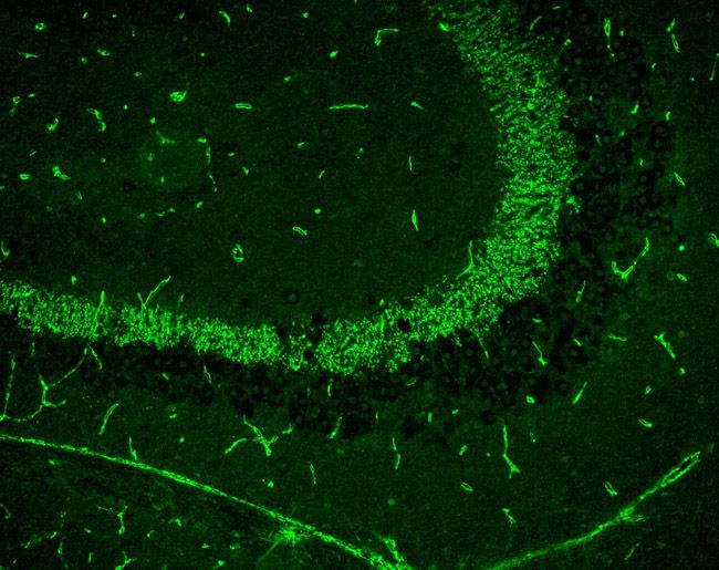
Immunofluorescent staining of mouse brain mosssy fiber terminals using Rb anti-ZO-1 (Mid) (Cat. No. 40-2200). Image courtesy of James I. Nagy, PhD, University of Manitoba, Canada.
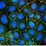
Indirect immunofluorescence staining of MDCKII cells using Zymed Rb anti-ZO-1 (Mid) (Cat. No. 40-2200). Image courtesy of Jacey Bennis and Dr. James Anderson, University of North Carolina at Chapel Hill, NC.
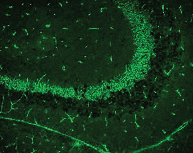
Immunofluorescent staining of mouse brain mosssy fiber terminals using Rb anti-ZO-1 (Mid) (Cat. no. 40-2200). Image courtesy of James I. Nagy, PhD, University of Manitoba, Canada.
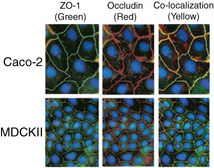
Indirect immunofluorescence staining of Caco-2 cells using Rb anti-ZO-1 (Mid). (PAD: ZMD.436) (Cat. No. 402200 (green), and anti-Occludin (red). Co-localization is in yellow. Image courtesy of Jacey Bennis and Dr. James Anderson, University of North Carolina at Chapel Hill, NC.
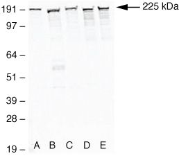
Western blot analysis of (A) MDCKII, (B) A431, (C) Caco-2, (D) Rat-1, and (E) NRK-52E cell lysates using Rb anti-ZO-1 (Mid) (Cat. no. 40-2200).


