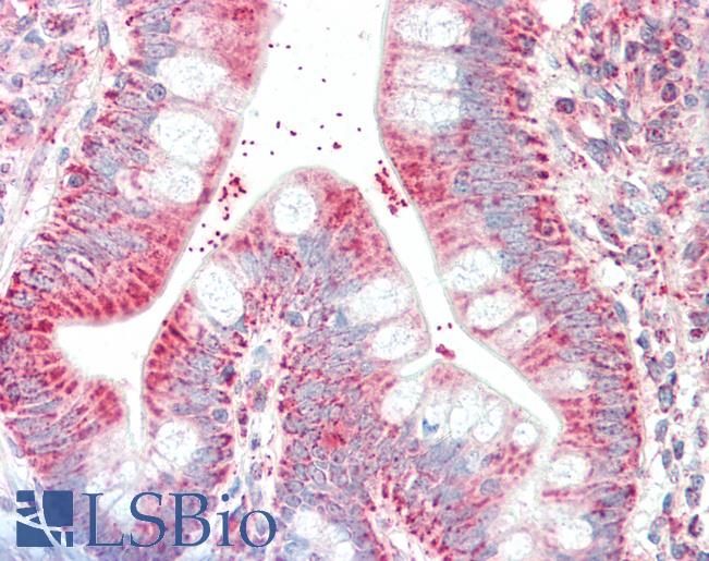
Anti-CSNK1A1 / CK1 Alpha antibody IHC staining of human small intestine. Immunohistochemistry of formalin-fixed, paraffin-embedded tissue after heat-induced antigen retrieval. Antibody LS-B10375 dilution 1:500.

High power image of HeLa cells stained with chicken casein kinase 1 alpha antibody (red) and our panspecific mouse monoclonal antibody to nuclear pore complexes MCA-39C7 (green). Casein kinase 1 alpha has a particulate distribution both in the nucleus and the cytoplasm. Blue is the Hoeschst DNA stain. High quality immunocytochemical images made using this antibody are shown in reference 4.

Western blot of whole rat spinal cord homogenate stained with chicken casein kinase 1 alpha antibody, at dilution of 1:10,000. A prominent band running with an apparent SDS-PAGE molecular weight of ~38kDa and a less prominent band at ~34kDa corresponds to other CK1 alpha isotypes.


