Immunocytochemistry:
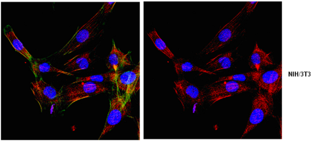
Representative lot data.
Confocal fluorescent analysis of NIH/3T3 cells using ABT60 (Red). Actin filaments have been labeled with AlexaFluor®488 -Phalloidin (Green). Nucleus is stained with DAPI (Blue). This antibody positively stains cytoplasm and membrane. The staining pattern is as expected: stains the membrane/cytoplasm, including ER, Golgi, etc,. Staining is also localized to a perinuclear compartment near the microtubule-organizing center (MTOC). Staining is also detected in tubulo-vesicular structures in the cell periphery that frequently localized along microtubules.

Immunohistochemistry Analysis:
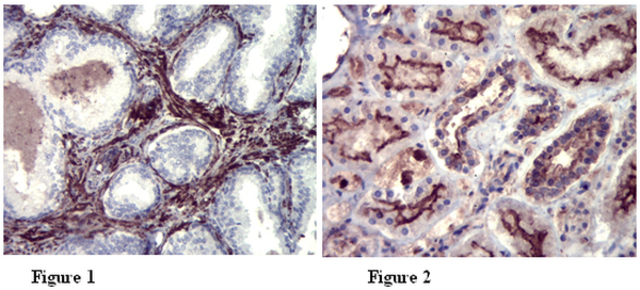
Representative lot data.
Paraffin-embedded fibromuscular stroma of human prostate (figure 1) and distal and proximal convoluted tubules in human kidney (figure 2) tissues were prepared using heat-induced epitope retrieval in citrate buffer, pH 6.0. Immunostaining was performed using a 1:500 dilution of Cat. No. ABT60, Anti-Whamm. Reactivity was detected using the IHC-Select® Detection Kit (Cat. No. DAB050). Positive cytoplasmic staining.

