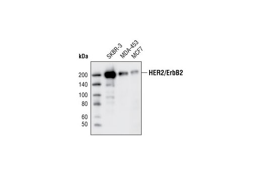
Western blot analysis of cell extracts from various cell lines, using HER2/ErbB2 (29D8) Rabbit mAb.

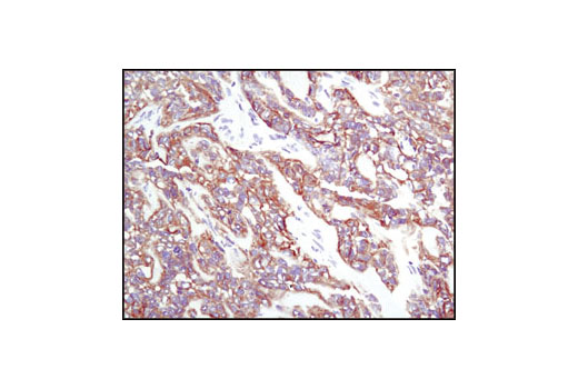
Immunohistochemical analysis of paraffin-embedded human breast carcinoma, using HER2/ErbB2 (29D8) Rabbit mAb.

Immunohistochemical analysis of paraffin-embedded human breast carcinoma using HER2/ErbB2 (29D8) RmAb in the presence of control peptide (left) or HER2/ErbB2 Blocking Peptide #1059 (right).

Immunohistochemical analysis of paraffin-embedded SKBr3 (high HER2) (left), MDA-MB-453 (moderate HER2) (middle) and MDA-MB-468 (low HER2) (right), using HER2/ErbB2 (29D8) Rabbit mAb.

Immunohistochemical analysis of frozen SKOV-3 xenograft using HER2/ErbB2 (29D8) Rabbit mAb.

Confocal immunofluorescent analysis of MDA-MB-453 cells (left) and MDA-MB-231 cells (right), using HER2/ErbB2 (29D8) Rabbit mAb (green). Blue pseudocolor = DRAQ5® #4084 (fluorescent DNA dye).

Flow cytometric analysis of MCF7 cells using HER2/ErbB2 (29D8) Rabbit mAb (blue) compared to a nonspecific negative control antibody (red).


