Western blot analysis of extracts from MCF7 (HER3+) and MDA-MB-231 (HER3-) cells using HER3/ErbB3 (D22C5) XP ® Rabbit mAb (upper) or β-Actin (D6A8) Rabbit mAb #8457 (lower).

Immunohistochemical analysis of paraffin-embedded non-small cell lung carcinoma using HER3/ErbB3 (D22C5) XP ® Rabbit mAb.

Immunohistochemical analysis of paraffin-embedded breast carcinoma using HER3/ErbB3 (D22C5) XP ® Rabbit mAb.

Immunohistochemical analysis of paraffin-embedded 293T cell pellets transfected with human erbB family members HER3, EGFR, HER2 and HER4 (from left to right as indicated) using HER3/ErbB3 (D22C5) XP ® Rabbit mAb (top panels). Transfections were confirmed using EGF Receptor (D38B1) XP ® Rabbit mAb #4267 (lower left), HER2/ErbB2 (D8F12) XP ® Rabbit mAb #4290 (lower middle) and HER4/ErbB4 (111B2) Rabbit mAb #4795 (lower right).

Immunohistochemical analysis of paraffin-embedded ovarian serous adenocarcinoma using HER3/ErbB3 (D22C5) XP ® Rabbit mAb.

Immunohistochemical analysis of paraffin-embedded MCF7 (left) or MDA-MB-231 (right) cell pellets using HER3/ErbB3 (D22C5) XP ® Rabbit mAb.

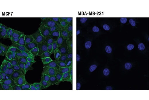
Confocal immunofluorescent analysis of MCF7 cells (HER3+, left) and MDA-MB-231 cells (HER-, right) using HER3/ErbB3 (D22C5) XP ® Rabbit mAb (green). Blue pseudocolor= DRAQ5 ® #4084 (fluorescent DNA dye).

Flow cytometric analysis of MCF7 cells using HER3/ErbB3 (D22C5) XP ® Rabbit mAb (blue) compared to Rabbit (DA1E) mAb IgG XP ® Isotype Control #3900. Anti-rabbit IgG (H+L), F(ab')2 fragment (Alexa Fluor ® 488 conjugate) #4412 was used as a secondary antibody.


