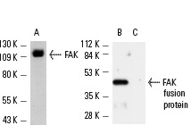
Western blot analysis of FAK expression in HeLa whole cell lysate (A). Western blot analysis of 20 ng of mouse recombinat FAK fusion protein (B) and an equal amount of an unrelated fusion protein (C). Antibodies tested include FAK (C-20)-G: sc-558-G (A) and FAK (C-20): sc-558 (B,C).
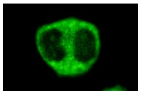
FAK (C-20): sc-558 Immunofluorescence staining of methanol-fixed HeLa cells showing cytoplasmic staining.
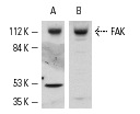
Western blot analysis of FAK expression in U-937 (A,B) whole cell lysates. Antibodies tested include FAK (C-20): sc-558 (A) and FAK (C-903): sc-932 (B).
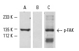
Western blot analysis of FAK phosphorylation in NIH/3T3 cells treated with anisomycin. Blots were probed with FAK (C-20): sc-558 (A), p-FAK (Ser 722)-R: sc-16662-R preincubated with its cognate phosphorylated peptide (B) and p-FAK (Ser 722)-R: sc-16662-R pre-incubated with its cognate non-phosphorylated peptide (C).
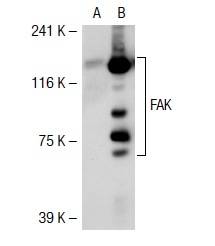
FAK (C-20): sc-558. Western blot analysis of FAK expression in non-transfected: sc-117752 (A) and human FAK transfected: sc-114600 (B) 293T whole cell lysates.
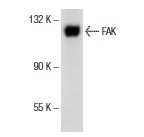
FAK (C-20)-G: sc-558-G. Western blot analysis of FAK expression in HeLa whole cell lysate.
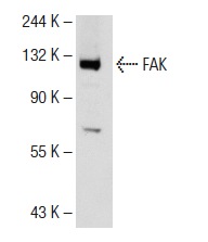
FAK (C-20): sc-558. Western blot analysis of FAK expression in HeLa whole cell lysate.
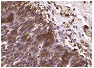
FAK (C-20): sc-558. Immunoperoxidase staining of formalin fixed, paraffin-embedded human urinary bladder tissue showing cytoplasmic staining of urothelial cells.
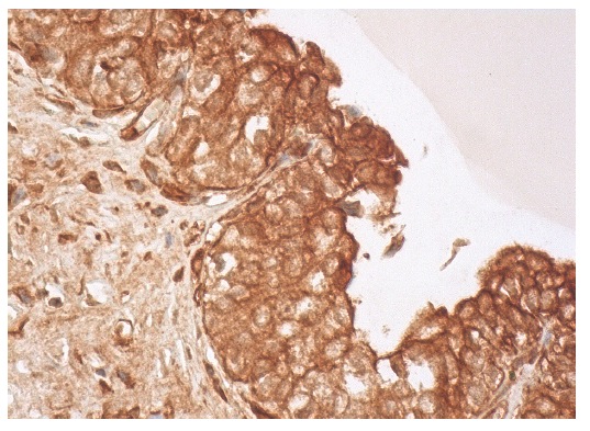
FAK (C-20)-G: sc-558-G. Immunoperoxidase staining of formalin fixed, paraffin-embedded human urinary bladder tissue showing cytoplasmic and membrane staining of urothelial cells.


