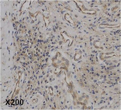
Immunohistochemical staining of IL-37 in human rheumatoid arthritis tissue. . Method: Immunohistochemical analysis of a paraffin-embedded rheumatoid arthritis tissue, showing endothelial cells and plasma cells (mature B cells) stained by anti-IL-37 at dilution 1:50 (under X200 lens, Brown color).

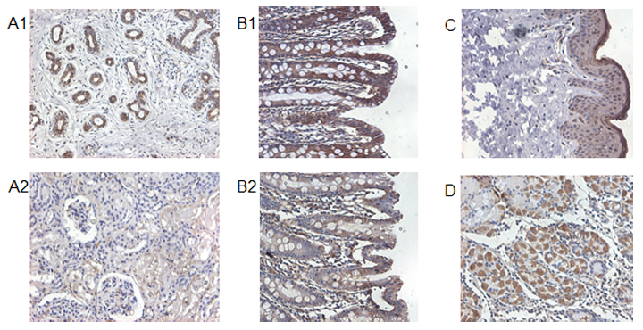
Immunohistochemical staining of IL-37 in human kidney (A1), human colon (B1), human skin (C) and human stomach (D) using anti-IL-37 . Human kidney (A2) and human colon (B2) are stained with negative control anti-rabbit IgG.

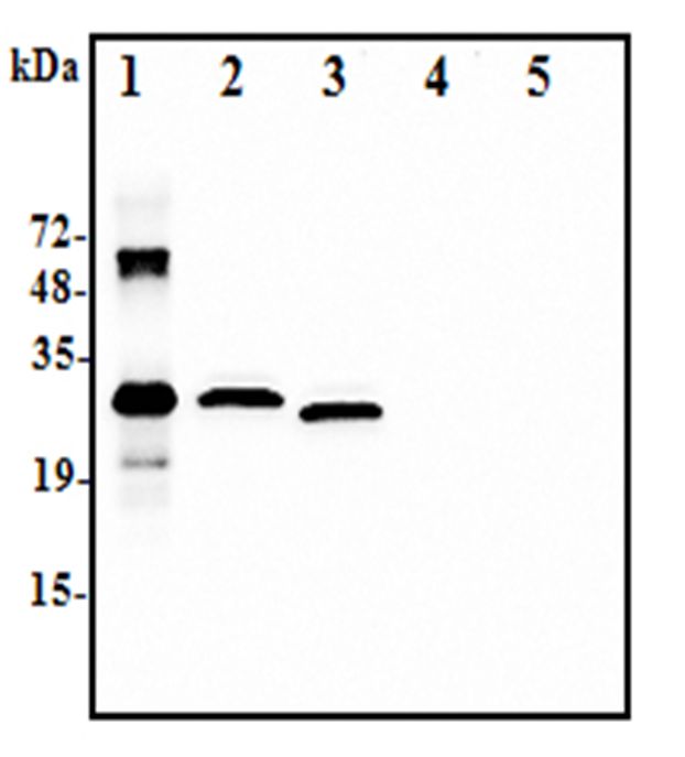
Western blot analysis using anti-IL-37 (human), pAb at 1:2.000 dilution:.1. Recombinant human IL-37-His (50ng).2. Human IL-37-FLAG transfected HEK293 cell lysate(100microg).3. Human IL-37-tag free transfected HEK293 cell lysate(100microg).4. Empty vector transfected HEK293 cell lysate(100microg).5. A unrelated protein-His (50ng)

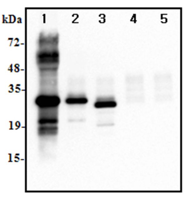
Immunoprecipitation of human IL-37 using anti-IL-37 at 1:500 dilutions:.1. Recombinant human IL-37-His (1microg).2. Human IL-37-FLAG transfected HEK293 cell lysate(500microg).3. Human IL-37-tag free transfected HEK293 cell lysate(500microg).4. Empty vector transfected HEK293 cell lysate(500microg).5. A unrelated protein-His (1microg)


