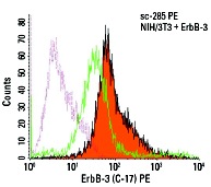
ErbB-3 (C-17) PE: sc-285 PE. Intracellular FCM analysis of fixed and permeabilized control (green line histogram) and ErbB-3 transfected (solid orange histogram) NIH/3T3 cells. Dotted pink histogram represents the isotype control, normal rabbit IgG: sc-3871.
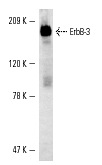
ErbB-3 (C-17)-G: sc-285-G. Western blot analysis of ErbB-3 transfected NIH/3T3 cells.
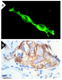
ErbB-3 (C-17): sc-285. Immunofluorescence staining of methanol-fixed NIH/3T3 cells transfected with ErbB-3 showing membrane localization (A). Immunoperoxidase staining of formalin-fixed, paraffin-embedded human breast carcinoma tissue showing membrane and cytoplasmic staining of epithelial cells (B).
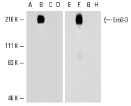
Western blot analysis of ErbB receptor expression in NIH/3T3 transfectants expressing between 10
5 and 10
6 molecules/cell of ErbB-2 (A,E), ErbB-3 (B,F) and ErbB-4 (C,G) and in NIH/3T3 (D,H) whole cell lysates. Antibodies tested include ErbB-3 (G-4): sc-7390 (A-D) and ErbB-3 (C-17): sc-285 (E-H).
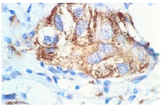
ErbB-3 (C-17): sc-285. Immunoperoxidase staining of formalin-fixed, paraffin-embedded human breast carcinoma tissue showing membrane and cytoplasmic staining of epithelial cells.
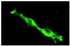
ErbB-3 (C-17): sc-285. Immunofluorescence staining of methanol-fixed NIH/3T3 cells transfected with ErbB-3 showing membrane localization.
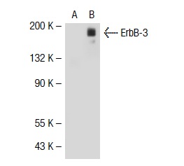
ErbB-3 (C-17)-G: sc-285-G. Western blot analysis of ErbB-3 expression in non-transfected: sc-117752 (A) and human ErbB-3 transfected: sc-111418 (B) 293T whole cell lysates.
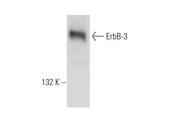
ErbB-3 (C-17)-G: sc-285-G. Western blot analysis of ErbB-3 expression in T-47D whole cell lysate.
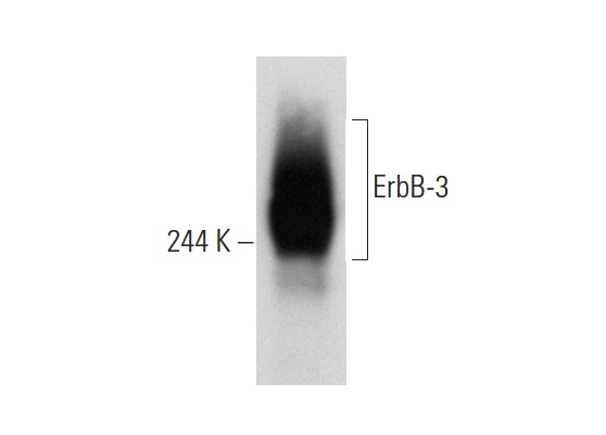
ErbB-3 (C-17): sc-285. Western blot analysis of ErbB-3 expression in T-47D whole cell lysate.


