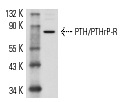
PTH/PTHrP-R (3D1.1): sc-12722. Western blot analysis of PTH/PTHrP-R expression in Saos-2 whole cell lysate.
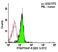
PTH/PTHrP-R (3D1.1) FITC: sc-12722 FITC. FCM analysis of human peripheral blood leukocytes. Black line histogram represents the isotype control, normal mouse IgG
1: sc-2855.
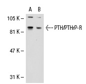
PTH/PTHrP-R (3D1.1): sc-12722. Western blot analysis of PTH/PTHrP-R expression in non-transfected: sc-117752 (A) and human PTH/PTHrP-R transfected: sc-114269 (B) 293T whole cell lysates.
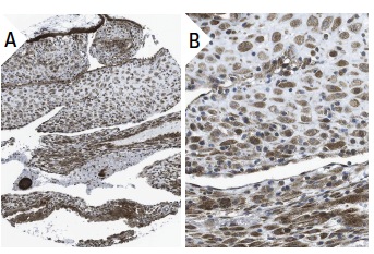
PTH/PTHrP-R (3D1.1): sc-12722. Immunoperoxidase staining of formalin fixed, paraffin-embedded human placenta tissue showing cytoplasmic staining of decidual and trophoblastic cells (low and high magnification). Kindly provided by The Swedish Human Protein Atlas (HPA) program.
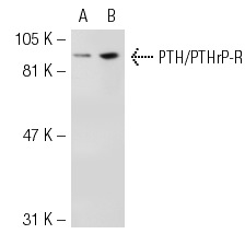
PTH/PTHrP-R (3D1.1): sc-12722. Western blot analysis of PTH1R expression in non-transfected: sc-117752 (A) and human PTH1R transfected: sc-112476 (B) 293T whole cell lysates.
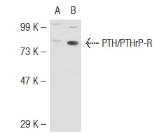
PTH/PTHrP-R (3D1.1): sc-12722. Western blot analysis of PTH/PTHrP-R expression in non-transfected: sc-117752 (A) and mouse PTH/PTHrP-R transfected: sc-122836 (B) 293T whole cell lysates.
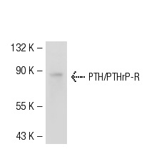
PTH/PTHrP-R (3D1.1): sc-12722. Western blot analysis of PTH/PTHrP-R expression in Caki-1 whole cell lysate.
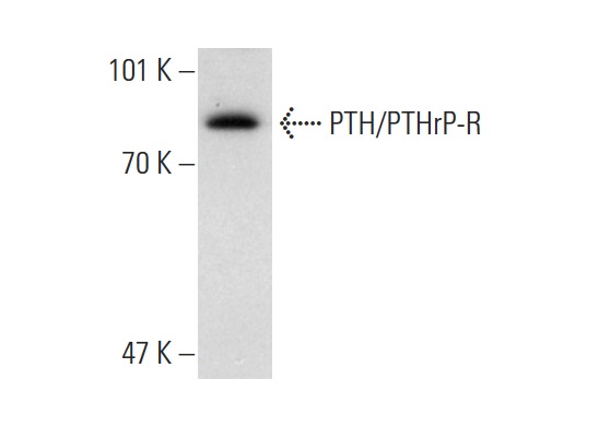
PTH/PTHrP-R (3D1.1): sc-12722. Western blot analysis of PTH/PTHrP-R expression in Caco-2 whole cell lysate.
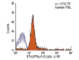
PTH/PTHrP-R (3D1.1) PE: sc-12722 PE. FCM analysis of human peripheral blood leukocytes. Black line histogram represents the isotype control, normal mouse IgG
1: sc-2866.
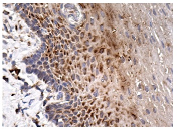
PTH/PTHrP-R (3D1.1): sc-12722. Immunoperoxidase staining of formalin fixed, paraffin-embedded human esophagus tissue showing cytoplasmic staining of squamous epithelial cells.


