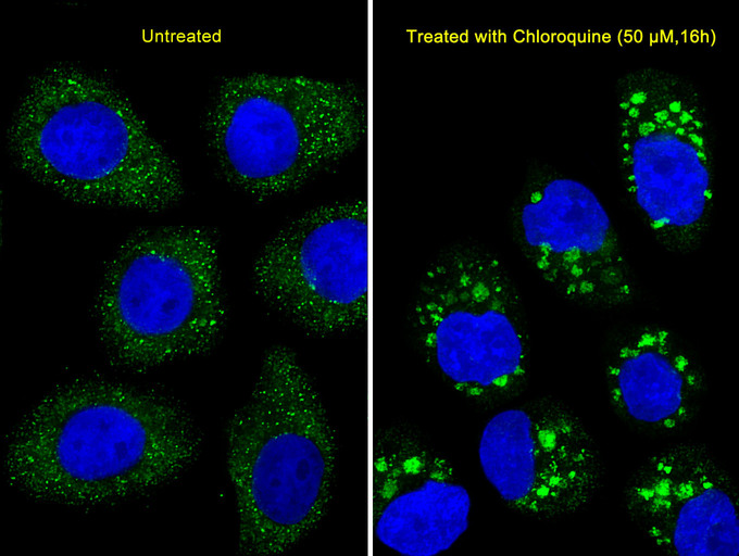
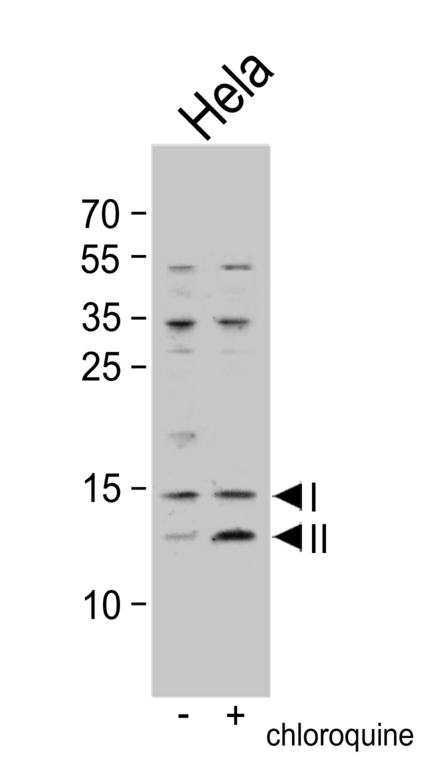
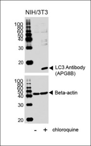
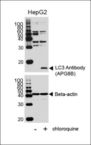
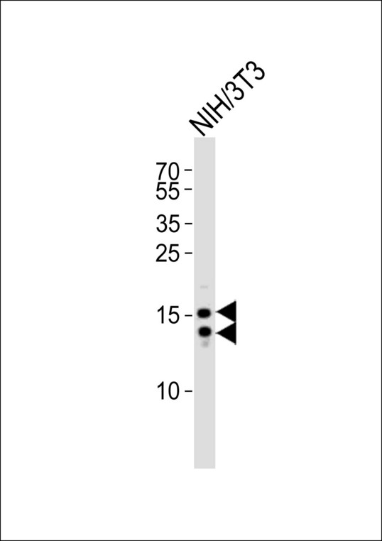
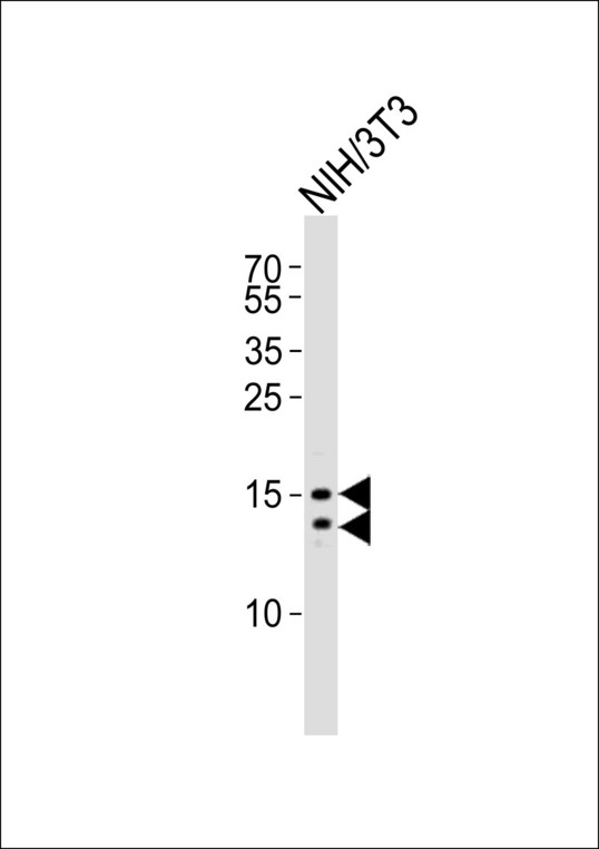
Anti-MAP1LC3B / LC3B Antibody (aa1-30)
| Name | Anti-MAP1LC3B / LC3B Antibody (aa1-30) |
|---|---|
| Supplier | LifeSpan Bioscience |
| Catalog | LS-C165711 |
| Prices | $295.00 |
| Sizes | 400 µl |
| Host | Rabbit |
| Clonality | Polyclonal |
| Applications | IHC IHC-P WB IP |
| Species Reactivities | Human, Rat, Zebrafish |
| Purity/Format | Protein A purified |
| Blocking Peptide | MAP1LC3B / LC3B Antibody Blocking Peptide |
| Description | Rabbit Polyclonal |
| Gene | MAP1LC3B |
| Conjugate | Unconjugated |
| Supplier Page | Shop |
Product images
Product References
Active ras triggers death in glioblastoma cells through hyperstimulation of - Active ras triggers death in glioblastoma cells through hyperstimulation of
Overmeyer JH, Kaul A, Johnson EE, Maltese WA. Mol Cancer Res. 2008 Jun;6(6):965-77.
Rab5 and class III phosphoinositide 3-kinase Vps34 are involved in hepatitis C - Rab5 and class III phosphoinositide 3-kinase Vps34 are involved in hepatitis C
Su WC, Chao TC, Huang YL, Weng SC, Jeng KS, Lai MM. J Virol. 2011 Oct;85(20):10561-71.
Caspase-6 activity in a BACHD mouse modulates steady-state levels of mutant - Caspase-6 activity in a BACHD mouse modulates steady-state levels of mutant
Gafni J, Papanikolaou T, Degiacomo F, Holcomb J, Chen S, Menalled L, Kudwa A, Fitzpatrick J, Miller S, Ramboz S, Tuunanen PI, Lehtimaki KK, Yang XW, Park L, Kwak S, Howland D, Park H, Ellerby LM. J Neurosci. 2012 May 30;32(22):7454-65.
