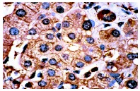
pan 14-3-3 (K-19): sc-629. Immunoperoxidase staining of formalin-fixed, paraffin-embedded human liver tumor showing cytoplasmic staining of hepatocytes.
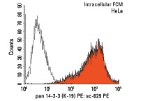
pan 14-3-3 (K-19) PE: sc-629 PE. Intracellular FCM analysis of fixed and permeabilized HeLa cells. Black line histogram represents the isotype control, normal rabbit IgG: sc-3871.
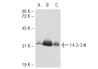
pan 14-3-3 (K-19): sc-629. Western blot analysis of 14-3-3 θ expression in non-transfected 293T: sc-117752 (A), mouse 14-3-3 θ transfected 293T: sc-117812 (B) and NIH/3T3 (C) whole cell lysates.
pan 14-3-3 (K-19): sc-629. Western blot analysis of 14-3-3 σ expression in non-transfected: sc-117725 (A) and human 14-3-3 σ transfected: sc-113566 (B) 293T whole cell lysates.
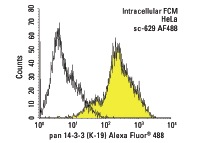
pan 14-3-3 (K-19) Alexa Fluor 488: sc-629 AF488. Intracellular FCM analysis of fixed and permeabilized HeLa cells. Black line histogram represents the isotype control, normal rabbit IgG: sc-45068.
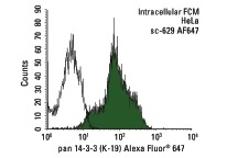
pan 14-3-3 (K-19) Alexa Fluor 647: sc-629 AF647. Intracellular FCM analysis of fixed and permeabilized HeLa cells. Black line histogram represents the isotype control, normal rabbit IgG: sc-24647.
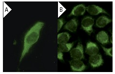
pan 14-3-3 (K-19): sc-629. Immunofluorescence staining of methanol-fixed NIH/3T3 cells showing cytoplasmic localization using indirect FITC (A) staining and HeLA cells using direct Alexa Fluor 488 (B) staining.
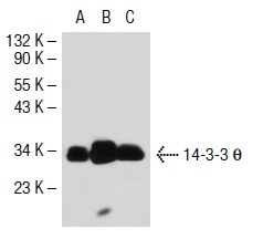
pan 14-3-3 (K-19): sc-629. Western blot analysis of pan 14-3-3 θ non-transfected 293T: sc-117752 (A), human pan 14-3-3 θ transfected 293T: sc-116633 (B) and NIH/3T3 (C) whole cell lysate.
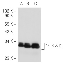
pan 14-3-3 (K-19): sc-629. Western blot analysis of 14-3-3 ζ expression in non-transfected 293T: sc-117752 (A), human 14-3-3 ζ transfected 293T: sc-111415 (B) and KNRK (C) whole cell lysates.
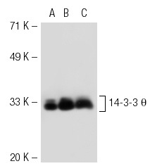
pan 14-3-3 (K-19): sc-629. Western blot analysis of 14-3-3 θ expression in non-transfected 293T: sc-117752 (A), human 14-3-3 θ transfected 293T: sc-111375 (B) and NIH/3T3 (C) whole cell lysates.
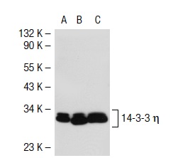
pan 14-3-3 (K-19): sc-629. Western blot analysis of 14-3-3 η expression in non-transfected 293T: sc-117752 (A), mouse 14-3-3 η transfected 293T: sc-117813 (B) and NIH/3T3 (C) whole cell lysates.
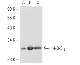
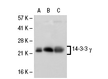
pan 14-3-3 (K-19): sc-629. Western blot analysis of 14-3-3 γ expression in non-transfected 293T: sc-117752 (A), human 14-3-3 γ transfected 293T: sc-113231 (B) and A-431 (C) whole cell lysates.
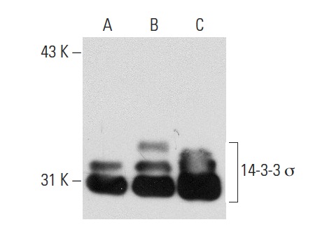
pan 14-3-3 (K-19): sc-629. Western blot analysis of 14-3-3 σ expression in non-transfected 293T: sc-117752 (A), human 14-3-3 σ transfected 293T sc-175742 (B) and HeLa (C) whole cell lysates.
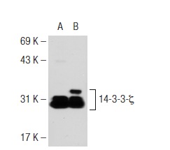
pan 14-3-3 (K-19): sc-629. Western blot analysis of 14-3-3 ζ expression in non-transfected: sc-110760 (A) and human 14-3-3 ζ transfected: sc-175740 (B) 293 whole cell lysates.
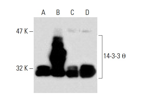
pan 14-3-3 (K-19): sc-629. Western blot analysis of 14-3-3 θ expression in non-transfected 293T: sc-117752 (A), human 14-3-3 θ transfected 293T: sc-127856 (B) and PC-12 (C) whole cell lysates and mouse placenta tissue extract (D).
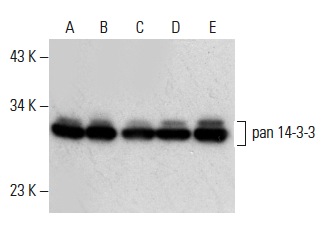
pan 14-3-3 (K-19)-G: sc-629-G. Western blot analysis of pan 14-3-3 expression in A-431 (A), K-562 (B), U-937 (C), HeLa (D) and Jurkat (E) whole cell lysates.
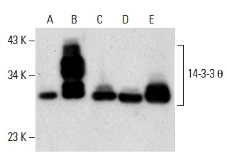
pan 14-3-3 (K-19)-G: sc-629-G. Western blot analysis of 14-3-3 θ expression in non-transfected 293T: sc-117752 (A), human 14-3-3 θtransfected 293T: sc-127856 (B), SK-N-MC (C) and PC-12 (D) whole cell lysates and mouse cerebellum tissue extract (E).
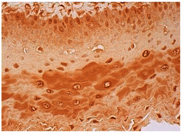
pan 14-3-3 (K-19): sc-629. Immunoperoxidase staining of formalin fixed, paraffin-embedded human esophagus tissue showing cytoplasmic and nuclear staining of squamous epithelial cells.


