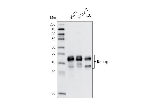
Western blot analysis of extracts from NCCIT, NTERA-2 and iPS cells using Nanog (D73G4) XP ® Rabbit mAb.
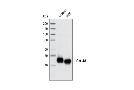
Western blot analysis of extracts from NTERA2 and mouse embryonic stem cells (mESCs) using Oct-4A (C30A3) Rabbit mAb.
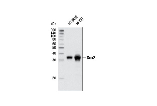
Western blot analysis of extracts from NTERA2 and NCCIT cells using Sox2 (D6D9) XP ® Rabbit mAb.
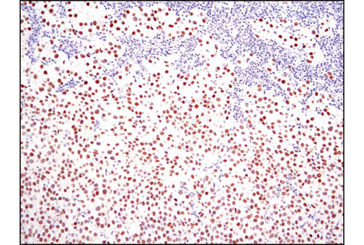
Immunohistochemical analysis of paraffin-embedded human seminoma using Nanog (D73G4) XP ® Rabbit mAb.
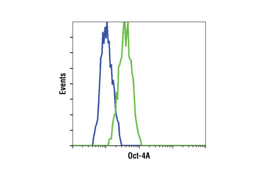
Flow cytometric analysis of HeLa cells (blue) and NCCIT cells (green) using Oct-4A (C30A3) Rabbit mAb.
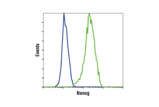
Flow cytometric analysis of HeLa cells (blue) and NTERA-2 cells (green) using Nanog (D73G4) XP ® Rabbit mAb.
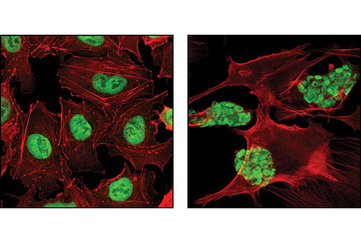
Confocal immunofluorescent analysis of NTERA2 (left) and mouse embryonic stem (mES) cells growing on mouse embryonic fibroblast (MEF) feeder cells (right) using Oct-4A (C30A3) Rabbit mAb (green). Actin filaments have been labeled with DY-554 phalloidin (red).
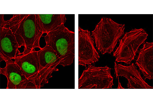
Confocal immunofluorescent analysis of NTERA-2 cells (left) and HeLa cells (right) using Nanog (D73G4) XP ® Rabbit mAb (green). Actin filaments have been labeled with DY-554 phalloidin (red).
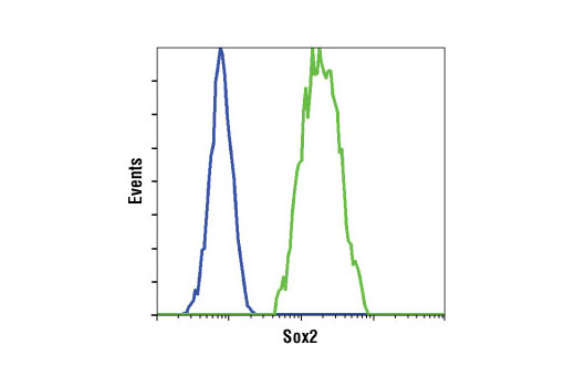
Flow cytometric analysis of HeLa cells (blue) and NTERA2 cells (green) using Sox2 (D6D9) XP ® Rabbit mAb.
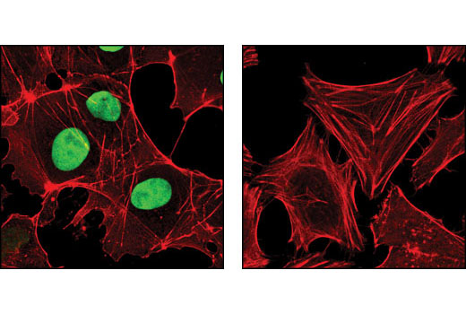
Confocal immunofluorescent analysis of NTERA2 (left) and HeLa (right) cells using Sox2 (D6D9) XP ® Rabbit mAb (green). Actin filaments have been labeled with DY-554 phalloidin (red).


