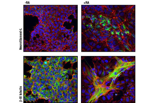
Confocal immunofluorescent analysis of NTERA-2 cells, untreated (left panel) or retinoic acid-treated (10 µM all-trans RA for 5 days) (right panel), using Neurofilament-L (C28E10) Rabbit mAb #2837 (green, upper), and β3-Tubulin (TU-20) Mouse mAb #4466 (green, lower). Actin filaments have been labeled with DY-554 phalloidin (red). Blue pseudocolor = DRAQ5 ® #4084 (fluorescent DNA dye). Note the appearance of neuronal markers and structures as cells differentiate along the neuronal lineage with retinoic acid treatment.

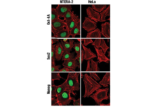
Confocal immunofluorescent analysis of NTERA-2 (left) and HeLa cells (right) using Oct-4A (C30A3) Rabbit mAb (green, upper), Sox2 (D6D9) XP® Rabbit mAb (green, middle) and Nanog (D73G4) XP® Rabbit mAb (green, lower).

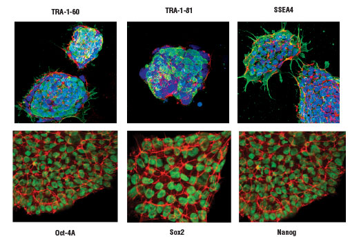
Projected confocal z-stack of human iPS cells using TRA-1-60(S) (TRA-1-60(S)) Mouse mAb (green, upper left), TRA-1-81 (TRA-1-81) Mouse mAb (green, upper middle), SSEA4 (MC813) Mouse mAb (green, upper right), Oct-4A (C30A3) Rabbit mAb (green, lower left), Sox2 (D6D9) XP ® Rabbit mAb (green, lower middle) and Nanog (D73G4) XP® Rabbit mAb (green, lower right). Actin filaments were labeled with DY-554 phalloidin (red). Blue pseudocolor = DRAQ5 ® #4084 (fluorescent DNA dye).

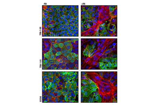
Confocal immunofluorescent analysis of NTERA-2 cells, untreated (left panel) or retinoic acid-treated (10 µM all-trans RA for 5 days) (right panel), using TRA-1-60(S) (TRA-1-60(S)) Mouse mAb (green, upper), TRA-1-81 (TRA-1-81) Mouse mAb (green, middle) and SSEA4 (MC813) Mouse mAb (green, lower). Actin filaments have been labeled with DY-554 phalloidin (red). Blue pseudocolor = DRAQ5 ® #4084 (fluorescent DNA dye). Note the loss of pluripotency markers (green) as cells differentiate along the neuronal lineage with retinoic acid treatment.


