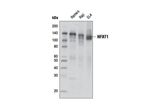
Western blot analysis of extracts from Ramos, Raji, and EL4 cells using NFAT1 (D43B1) XP ® Rabbit mAb.

Immunohistochemical analysis of paraffin-embedded human breast carcinoma using NFAT1 (D43B1) XP ® Rabbit mAb.

Immunohistochemical analysis of paraffin-embedded human colon carcinoma using NFAT1 (D43B1) XP ® Rabbit mAb.

Immunohistochemical analysis of paraffin-embedded human T-cell lymphoma using NFAT1 (D43B1) XP ® Rabbit mAb.

Immunohistochemical analysis of paraffin-embedded cell pellets, Jurkat (left) or LNCaP (right), using NFAT1 (D43B1) XP ® Rabbit mAb.

Confocal immunofluorescent analysis of MCF7 cells, untreated (left) or treated with Ionomycin #9995 (1 μM, 1 hr; right), using NFAT1 (D43B1) XP ® Rabbit mAb (green). Actin filaments were labeled with DY-554 phalloidin (red). Blue pseudocolor = DRAQ5 ® #4084 (fluorescent DNA dye).

Flow cytometric analysis of HCT 116 using NFAT1 (D43B1) XP ® Rabbit mAb (blue) compared to concentration matched Rabbit (DA1E) mAb IgG XP ® Isotype Control #3900 (red).


