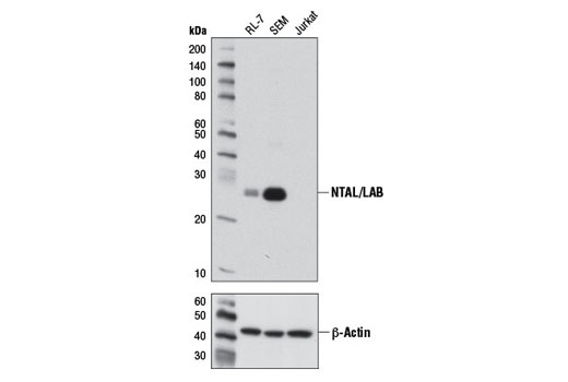
Western blot analysis of extracts from RL-7, SEM, and Jurkat cells using NTAL/LAB (D7I2B) Rabbit mAb (upper) or β-Actin (D6A8) Rabbit mAb #8457 (lower).

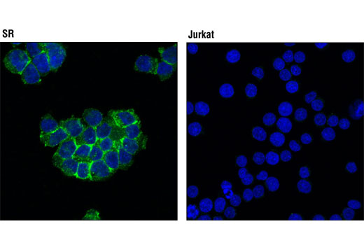
Confocal immunofluorescent analysis of SR (positive; left) and Jurkat (negative; right) cells using NTAL/LAB (D7I2B) Rabbit mAb (green). Blue pseudocolor = DRAQ5 ® #4084 (fluorescent DNA dye).

Human whole blood was fixed, lysed, and permeabilized as per the Cell Signaling Technology Flow Alternate Protocol and stained using NTAL/LAB (D7I2B) Rabbit mAb. Samples were co-stained with CD19-APC and CD3-PE to identify B and T cell populations, respectively. The forward/side scatter lymphocyte gate and CD19 + B cell population gate were combined and applied to a histogram depicting the mean fluorescence intensity of NTAL/LAB (blue) and a concentration-matched isotype control (red). Anti-rabbit IgG (H+L), F(ab') 2 Fragment (Alexa Fluor ® 488 Conjugate) #4412 was used as a secondary antibody.


