Phospho-p53 (Ser15) (16G8) Mouse mAb
| Name | Phospho-p53 (Ser15) (16G8) Mouse mAb |
|---|---|
| Supplier | Cell Signaling Technology |
| Catalog | 9286 |
| Prices | $109.00, $287.00, $678.00 |
| Sizes | 20 µl (2 western blots), 100 µl (20 western blots), 300 µl (60 western blots) |
| Host | Mouse |
| Clonality | Monoclonal |
| Isotype | IgG1 |
| Clone | 16G8 |
| Applications | WB ICC/IF FC |
| Species Reactivities | Human |
| Antigen | Monoclonal antibody is produced by immunizing animals with a synthetic phosphopeptide corresponding to residues surrounding Ser15 of human p53. |
| Description | Mouse Monoclonal |
| Gene | TP53 |
| Supplier Page | Shop |
Product images
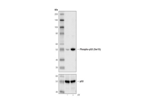 Western blot analysis of extracts from HT29 cells, untreated or UV-treated (100 mJ/cm2, 1 hr), using Phospho-p53 (Ser15) (16G8) Mouse mAb (upper) or p53 (DO-7) Mouse mAb #48818 (lower).
 Confocal immunofluorescent analysis of HT-29 cells, untreated (left) or UV-treated (right), using Phospho-p53 (Ser15) (16G8) Mouse mAb (green). Actin filaments have been labeled with Alexa Fluor® 555 phalloidin (red).
 Flow cytometric analysis of HT-29 cells, untreated (blue) or UV-treated (green), using Phospho-p53 (Ser15) (16G8) Mouse mAb compared to a nonspecific negative control antibody (red).