Western blot analysis of extracts from various cell lines using PDGF Receptor α (D13C6) XP ® Rabbit mAb (upper), PDGF Receptor β (28E1) Rabbit mAb #3169 (middle), and β-Actin Antibody #4967 (lower).

Immunohistochemical analysis of paraffin-embedded human astrocytoma using PDGF Receptor α (D13C6) XP ® Rabbit mAb.

Immunohistochemical analysis of paraffin-embedded NCI-H1703 (PDGFRα+, left) and HCC827 (PDGFRα-, right) cell pellets using PDGF Receptor α (D13C6) XP ® Rabbit mAb.

Immunohistochemical analysis of paraffin-embedded human leiomyosarcoma using PDGF Receptor α (D13C6) XP ® Rabbit mAb.

Confocal immunofluorescent analysis of NCI-H1703 (left), A172 (center) and HCC827 cells (right) using PDGF Receptor α (D13C6) XP ® Rabbit mAb (green). Actin filaments were labeled with DY-554 phalloidin (red). Blue pseudocolor = DRAQ5 ® #4084 (fluorescent DNA dye).

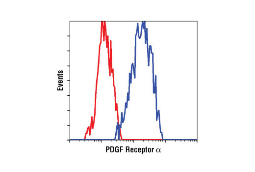
Flow cytometric analysis of A-204 cells using PDGF Receptor α (D13C6) XP ® Rabbit mAb (blue) compared to a nonspecific negative control antibody (red).


