PDGFRA

Gene Symbol PDGFRA
Entrez Gene 5156
Alt Symbol CD140A, PDGFR-2, PDGFR2, RHEPDGFRA
Species Human
Gene Type protein-coding
Description platelet-derived growth factor receptor, alpha polypeptide
Other Description CD140 antigen-like family member A|CD140a antigen|PDGF-R-alpha|PDGFRA/BCR fusion|alpha-type platelet-derived growth factor receptor|platelet-derived growth factor receptor 2|platelet-derived growth factor receptor alpha|rearranged-in-hypereosinophilia-platelet derived growth factor receptor alpha fusion protein
Swissprots E9PBH0 P16234 B2RE69 Q96KZ7 Q6P4H5 Q9UD28
Accessions BAA08742 CAJ33669 CAJ33686 CAN37625 CBH30545 EAX05457 EAX05458 P16234 AA599881 AA625689 AK308353 AK311006 AK316578 BAG38166 AV689272 BC015186 AAH15186 BC063414 AAH63414 DA678599 L25829 M21574 AAA96715 M22734 AAA60048 M30494 X76079 XM_005265743 XP_005265800 XM_006714039 XP_006714102 XM_006714041 XP_006714104 XM_011534385 XP_011532687 XM_011534386 XP_011532688 NM_006206 NP_006197
Function Tyrosine-protein kinase that acts as a cell-surface receptor for PDGFA, PDGFB and PDGFC and plays an essential role in the regulation of embryonic development, cell proliferation, survival and chemotaxis. Depending on the context, promotes or inhibits cell proliferation and cell migration. Plays an important role in the differentiation of bone marrow-derived mesenchymal stem cells. Required for normal skeleton development and cephalic closure during embryonic development. Required for normal development of the mucosa lining the gastrointestinal tract, and for recruitment of mesenchymal cells and normal development of intestinal villi. Plays a role in cell migration and chemotaxis in wound healing. Plays a role in platelet activation, secretion of agonists from platelet granules, and in thrombin-induced platelet aggregation. Binding of its cognate ligands - homodimeric PDGFA, homodimeric PDGFB, heterodimers formed by PDGFA and PDGFB or homodimeric PDGFC -leads to the activation of sever
Subcellular Location Cell membrane {ECO:0000269|PubMed:14644164, ECO:0000269|PubMed:2554309, ECO:0000269|PubMed:8188664}; Single- pass type I membrane protein {ECO:0000269|PubMed:14644164, ECO:0000269|PubMed:2554309, ECO:0000269|PubMed:8188664}. Note=The activated receptor is rapidly internalized and degraded.
Tissue Specificity Detected in platelets (at protein level). Widely expressed. Detected in brain, fibroblasts, smooth muscle, heart, and embryo. Expressed in primary and metastatic colon tumors and in normal colon tissue. {ECO:0000269|PubMed:2536956, ECO:0000269|PubMed:7896447, ECO:0000269|PubMed:8188664}.
Top Pathways Rap1 signaling pathway, MicroRNAs in cancer, Central carbon metabolism in cancer, PI3K-Akt signaling pathway, Choline metabolism in cancer

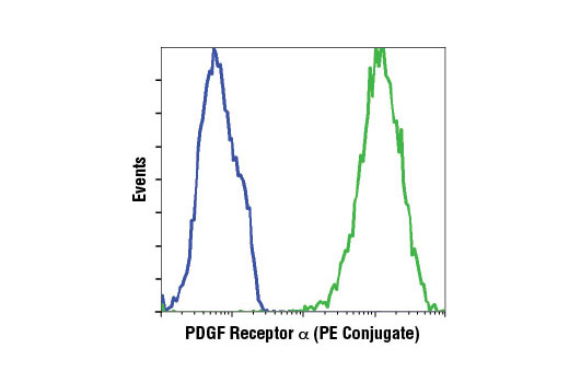
PDGF Receptor α (D13C6) XP ® Rabbit mAb (PE Conjugate) - 8533 from Cell Signaling Technology

Prices $329.00
Sizes 100 µl (50 tests)
Host Rabbit
Clonality Monoclonal
Clone D13C6
Flow cytometric analysis of HCC827 (blue) or NCI-H1703 (green) cells using PDGF Receptor α (D13C6) XP ® Rabbit mAb (PE Conjugate).

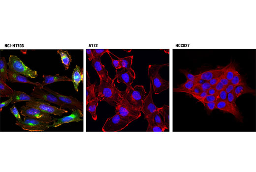
PDGF Receptor α (D13C6) XP ® Rabbit mAb (Alexa Fluor ® 594 Conjugate) - 9015 from Cell Signaling Technology

Prices $329.00
Sizes 100 µl (50 tests)
Host Rabbit
Clonality Monoclonal
Clone D13C6
Confocal immunofluorescent analysis of NCI-H1703 (α+/β-; left), A172 (α-/β+; center), and HCC827 (α-/β-; right) cells using PDGF Receptor α (D13C6) XP® Rabbit mAb (Alexa Fluor® 594 Conjugate) (red). Actin filaments were labeled with Alexa Fluor® 488 phalloidin (green). Blue pseudocolor = DRAQ5® #4084 (fluorescent DNA dye).

PDGF Receptor α (D13C6) XP ® Rabbit mAb (Biotinylated) - 5278 from Cell Signaling Technology

Prices $329.00
Sizes 100 µl (10 western blots)
Host Rabbit
Clonality Monoclonal
Clone D13C6
Western blot analysis of extracts from NCI-H1703, HCC827, and U-118 MG cells using PDGF Receptor α (D13C6) XP ® Rabbit mAb (Biotinylated).

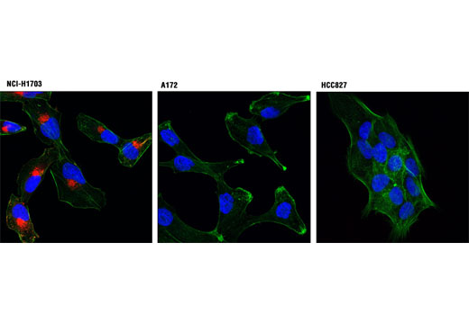
PDGF Receptor α (D13C6) XP ® Rabbit mAb (Alexa Fluor ® 488 Conjugate) - 8871 from Cell Signaling Technology

Prices $329.00
Sizes 100 µl (50 tests)
Host Rabbit
Clonality Monoclonal
Clone D13C6
Confocal immunofluorescent analysis of NCI-H1703 (α+/β-; left), A172 (α-/β+; center), and HCC827 (α-/β-; right) cells using PDGF Receptor α (D13C6) XP ® Rabbit mAb (Alexa Fluor ® 488 Conjugate) (green). Actin filaments were labeled with DY-554 phalloidin (red). Blue pseudocolor = DRAQ5 ® #4084 (fluorescent DNA dye).

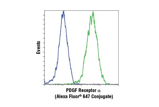
PDGF Receptor α (D13C6) XP ® Rabbit mAb (Alexa Fluor ® 647 Conjugate) - 5876 from Cell Signaling Technology

Prices $329.00
Sizes 100 µl (50 tests)
Host Rabbit
Clonality Monoclonal
Clone D13C6
Flow cytometric analysis of HCC827 (blue) or NCI-H1703 (green) cells using PDGF Receptor α (D13C6) XP ® Rabbit mAb (Alexa Fluor ® 647 Conjugate).

PDGF Receptor α (D13C6) XP ® Rabbit mAb (Alexa Fluor ® 555 Conjugate) - 8893 from Cell Signaling Technology

Prices $329.00
Sizes 100 µl (50 tests)
Host Rabbit
Clonality Monoclonal
Clone D13C6
Confocal immunofluorescent analysis of NCI-H1703 (α+/β-; left), A172 (α-/β+; center), and HCC827 (α-/β-; right) cells using PDGF Receptor α (D13C6) XP ® Rabbit mAb (Alexa Fluor ® 555 Conjugate) (red). Actin filaments were labeled with Alexa Fluor ® 488 phalloidin (green). Blue pseudocolor = DRAQ5 ® #4084 (fluorescent DNA dye).

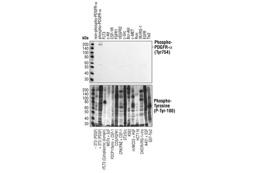
Phospho-PDGF Receptor α (Tyr754) (23B2) Rabbit mAb - 2992 from Cell Signaling Technology

Prices $109.00, $287.00
Sizes 20 µl (2 western blots), 100 µl (10 western blots)
Host Rabbit
Clonality Monoclonal
Clone 23B2
Phospho-PDGF Receptor α (Tyr754) (23B2) Rabbit mAb specifically binds to tyrosine phosphorylated PDGF receptor α, but not other phosphorylated tyrosine kinases. Western blot analysis of of extracts from cells expressing different activated tyrosine kinase proteins, using Phospho-PDGF Receptor α (Tyr754) (23B2) Rabbit mAb (upper) or Phospho-Tyrosine Mouse mAb (P-Tyr-100) #9411 (lower).

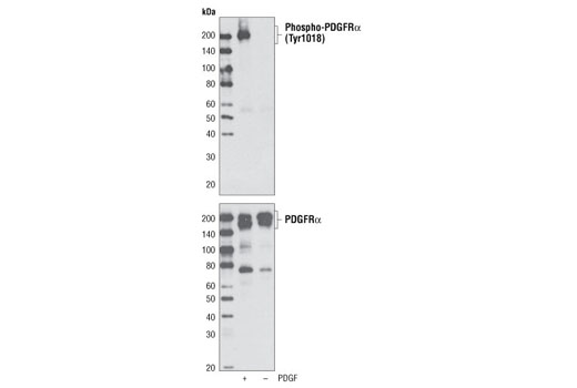
Phospho-PDGF Receptor α (Tyr1018) Antibody - 4547 from Cell Signaling Technology

Prices $109.00, $287.00
Sizes 20 µl (2 western blots), 100 µl (10 western blots)
Host Rabbit
Clonality Polyclonal
Western blot analysis of cell extracts from NIH/3T3 cells, untreated or treated with PDGF-AA, using Phospho-PDGF Receptor α (Tyr1018) Antibody (upper) or PDGF Receptor α Antibody #3164 (lower).

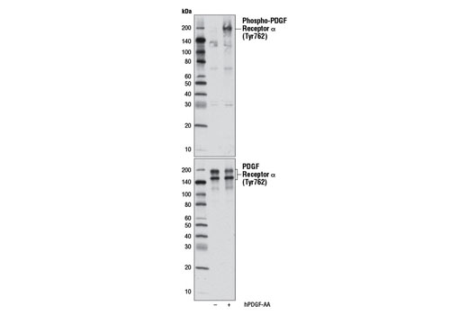
Phospho-PDGF Receptor α (Tyr762) Antibody - 12022 from Cell Signaling Technology

Prices $279.00
Sizes 100 µl (10 western blots)
Host Rabbit
Clonality Polyclonal
Western blot analysis of extracts from NCI-H1703 cells serum-starved overnight, untreated (-) or treated with Human Platelet-Derived Growth Factor AA (hPDGF-AA) #8913 (100 ng/ml, 10 min;+), using Phospho-PDGF Receptor α (Tyr762) Antibody (upper) or PDGF Receptor α (D13C6) XP® Rabbit mAb #5241 (lower).

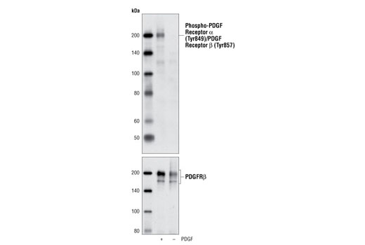
Phospho-PDGF Receptor α (Tyr849)/PDGF Receptor β (Tyr857) (C43E9) Rabbit mAb - 3170 from Cell Signaling Technology

Prices $109.00, $287.00
Sizes 20 µl (2 western blots), 100 µl (10 western blots)
Host Rabbit
Clonality Monoclonal
Clone C43E9
Western blot analysis of NIH/3T3 cells, untreated or treated with PDGFbb using Phospho-PDGF Receptor α (Tyr849)/PDGF Receptor β (Tyr857) (C43E9) Rabbit mAb (upper) and PDGF Receptor β (28E1) Rabbit mAb #3169 (lower).

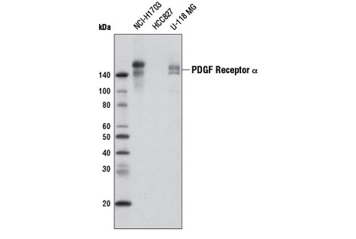
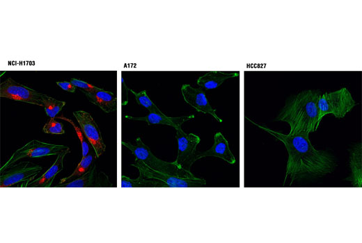
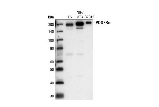
PDGF Receptor α (D13C6) XP ® Rabbit mAb - 5241 from Cell Signaling Technology

Prices $109.00, $277.00
Sizes 20 µl (2 western blots), 100 µl (10 western blots)
Host Rabbit
Clonality Monoclonal
Clone D13C6
Western blot analysis of extracts from various cell lines using PDGF Receptor α (D13C6) XP ® Rabbit mAb (upper), PDGF Receptor β (28E1) Rabbit mAb #3169 (middle), and β-Actin Antibody #4967 (lower).

PDGF Receptor α (D1E1E) XP ® Rabbit mAb - 3174 from Cell Signaling Technology

Prices $109.00, $277.00
Sizes 20 µl (2 western blots), 100 µl (10 western blots)
Host Rabbit
Clonality Monoclonal
Clone D1E1E
Western blot analysis of extracts from NIH/3T3 and human skeletal muscle cells (SKMC), untreated or treated with PDGF-BB, using PDGF Receptor α (D1E1E) XP ® Rabbit mAb.

PDGF Receptor α Antibody - 3164 from Cell Signaling Technology

Prices $246.00
Sizes 100 µl (10 western blots)
Host Rabbit
Clonality Polyclonal
Western blot analysis of extracts from L6, NIH/3T3 and C2C12 cells, using PDGF Receptor α Antibody.

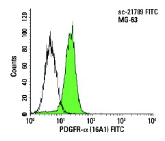
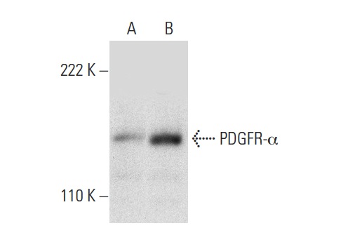
PDGFR-α (16A1) - sc-21789 from Santa Cruz Biotechnology

Prices $279.00
Sizes 200 µg/ml
Host Mouse
Clonality Monoclonal

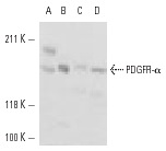
PDGFR-α (C-9) - sc-398206 from Santa Cruz Biotechnology

Prices $279.00
Sizes 200 µg/ml
Host Mouse
Clonality Monoclonal
PDGFR-α (C-9): sc-398206. Western blot analysis of PDGFR-α expression in COLO 320DM (A) and HeLa (B) whole cell lysates.

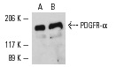
PDGFR-α (951) - sc-431 from Santa Cruz Biotechnology

Prices $279.00
Sizes 200 µg/ml
Host Rabbit
Clonality Polyclonal
PDGFR-α (951): sc-431. Western blot analysis of PDGFR-α expression in NIH/3T3 (A), CCD-1064Sk (B), U-2 OS (C) and Saos-2 (D) whole cell lysates.

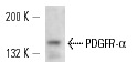
PDGFR-α (C-20) - sc-338 from Santa Cruz Biotechnology

Prices $279.00
Sizes 200 µg/ml
Host Rabbit
Clonality Polyclonal
PDGFR-α (C-20): sc-338. Western blot analysis of PDGFR-α expression in NIH/3T3 whole cell lysates (A,B).

PDGFR-α (V-17) - sc-31178 from Santa Cruz Biotechnology

Prices $279.00
Sizes 200 µg/ml
Host Goat
Clonality Polyclonal
PDGFR-α (V-17): sc-31178. Western blot analysis of PDGFR-α expression in HeLa whole cell lysate.

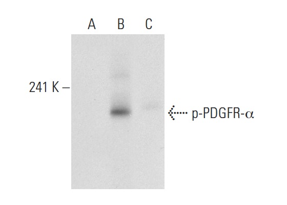
p-PDGFR-α (Tyr 720) - sc-12910 from Santa Cruz Biotechnology

Prices $279.00
Sizes 200 µg/ml
Host Goat
Clonality Polyclonal
p-PDGFR-α (Tyr 720)-R: sc-12910-R. Western blot analysis of PDGFR-α phosphorylation in untreated (A), PDGF treated (B) and PDGF and lambda protein phosphatase treated (C) NIH/3T3 whole cell lysates.

p-PDGFR-α (Tyr 754) - sc-12911 from Santa Cruz Biotechnology

Prices $279.00
Sizes 200 µg/ml
Host Goat
Clonality Polyclonal
p-PDGFR-α (Tyr 754): sc-12911. Immunoperoxidase staining of formalin-fixed, paraffin-embedded medulloblastoma tissue showing membrane and cytoplasmic staining. Kindly provided by Dr. Huizhen Zhang, Rood Laboratory, Children's National Medical Center.

Anti-PDGF receptor alpha + beta (phospho Y572 + Y574) antibody - ab5443 from Abcam

Prices $370.00
Sizes 50 µl
Host Rabbit
Clonality Polyclonal
Peptide Competition: Extracts prepared from NIH3T3 cells left unstimulated (1) and stimulated with PDGF (2-5) were resolved by SDS-PAGE on a 10% polyacrylamide gel and transferred to PVDF. Membranes were blocked with a 5% BSA-TBST buffer overnight at 4°C, then were incubated with 0.50 µg/mL ab5443 antibody for two hours at room temperature in a 1% BSA-TBST buffer, following prior incubation with: no peptide (1, 2), the non-phosphopeptide corresponding to the immunogen (3), a generic phosphotyrosine containing peptide (4), or, the phosphopeptide immunogen (5). After washing, membranes were incubated with goat F(ab’)2 anti-rabbit IgG alkaline phosphatase and bands were detected using the Tropix WesternStar method. The data show that only the peptide corresponding to ab5443 blocks the antibody signal, thereby demonstrating the specificity of the antibody.

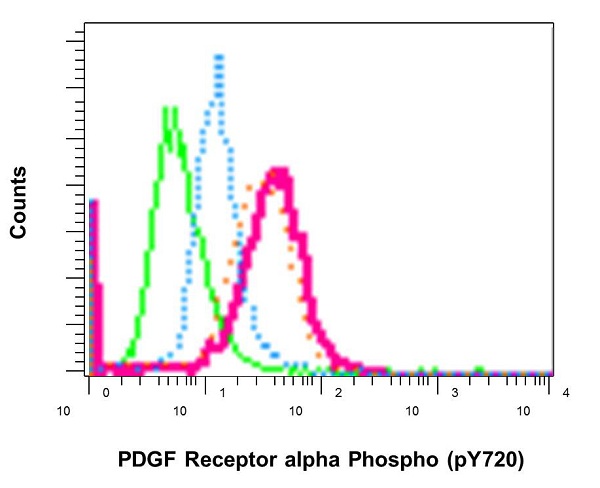
Anti-PDGF Receptor alpha (phospho Y720) antibody [EP2478] - ab134068 from Abcam

Prices $313.00
Sizes 100 µl
Host Rabbit
Clonality Monoclonal
Clone EP2478
Flow Cytometry analysis of permeabilized NIH/3T3 cells labelling PDGF Receptor alpha (phospho Y720) with ab134068. Green - untreated, Red - pervanadate-treated, Blue - preincubated with phospho-PDGF Receptor alpha (phospho Y720) peptide, Orange - preincubated with non-phospho-PDGR Receptor alpha peptide.

Anti-PDGF Receptor alpha (phospho Y742) antibody - ab5452 from Abcam

Prices $378.00
Sizes 50 µl
Host Rabbit
Clonality Polyclonal
Peptide Competition: Extracts prepared from NIH3T3 cells left unstimulated (1) and stimulated with PDGF (2-5) were resolved by SDS-PAGE on a 10% polyacrylamide gel and transferred to PVDF. Membranes were blocked with a 5% BSA-TBST buffer overnight at 4°C, then were incubated with 0.50 µg/mL ab5452 antibody for two hours at room temperature in a 1% BSA-TBST buffer, following prior incubation with: no peptide (1, 2), the nonphosphopeptide corresponding to the immunogen (3), a generic phosphotyrosine containing peptide (4), or, the phosphopeptide immunogen (5). After washing, membranes were incubated with goat F(ab’)2 anti-rabbit IgG alkaline phosphatase and bands were detected using the Tropix WesternStar method. The data show that only the peptide corresponding to ab5452 blocks the antibody signal, thereby demonstrating the specificity of the antibody.

Anti-PDGF Receptor alpha (phospho Y754) antibody - ab5460 from Abcam

Prices $394.00
Sizes 50 µl
Host Rabbit
Clonality Polyclonal
Peptide Competition: Extracts prepared from NIH3T3 cells left unstimulated (1) and stimulated with PDGF (2-5) were resolved by SDS-PAGE on a 10% polyacrylamide gel and transferred to PVDF. Membranes were blocked with a 5% BSA-TBST buffer overnight at 4°C, then were incubated with 0.50 µg/mL ab5460 antibody for two hours at room temperature in a 1% BSA-TBST buffer, following prior incubation with: no peptide (1, 2), the non-phosphopeptide corresponding to the immunogen (3), a generic phosphotyrosine containing peptide (4), or, the phosphopeptide immunogen (5). After washing, membranes were incubated with goat F(ab’)2 anti-rabbit IgG alkaline phosphatase and bands were detected using the Tropix WesternStar method. The data show that only the peptide corresponding to ab5460 blocks the antibody signal, thereby demonstrating the specificity of the antibody.

Anti-PDGF Receptor alpha (phospho Y849) antibody - ab79318 from Abcam

Prices $380.00
Sizes 100 µg
Host Rabbit
Clonality Polyclonal
All lanes : Anti-PDGF Receptor alpha (phospho Y849) antibody (ab79318) at 1/500 dilutionLane 1 : 293 cells extractLane 2 : 293 cells extract with Immunizing phosphopeptide at 5 µgLysates/proteins at 5 µg per lane.
Search more