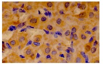
GRB7 (C-20): sc-606. Immunoperoxidase staining of formalin fixed, paraffin-embedded mouse ovary tissue showing membrane and cytoplasmic localization.
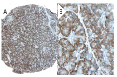
GRB7 (C-20): sc-606. Immunoperoxidase staining of formalin fixed, paraffin-embedded human pancreas tissue showing cytoplasmic staining of exocrine pancreas cells (low and high magni?fication). Kindly provided by The Swedish Human Protein Atlas (HPA) program.
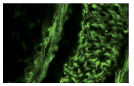
GRB7 (C-20): sc-606. Immunofluorescence staining of normal mouse intestine frozen section showing cytoplasmic staining.
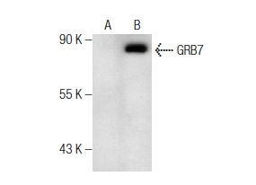
GRB7 (C-20): sc-606. Western blot analysis of GRB7 expression in A-431 (A) and EGF treated A-431 (B) whole cell lysates.