Anti-ROBO1 antibody IHC of human brain. Immunohistochemistry of formalin-fixed, paraffin-embedded tissue after heat-induced antigen retrieval. Antibody LS-B81 concentration 5 ug/ml.

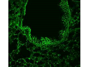
Immunofluorescence - Anti-ROBO-1 Antibody. 1/50 staining mouse lung tissue sections (adult, frozen 100mum whole mount sections) by IHC-Fr. The tissue was paraformaldehyde fixed and permeabilized with triton x-100 before incubation with the antibody for 16 hours at 4°C.

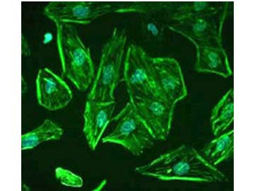
Immunofluorescence - Anti-ROBO-1 Antibody. Staining of ROBO1 in undifferentiated, immortalized human podocytes by Immunocytochemistry/ Immunofluorescence. Cells were fixed with 2% paraformaldehyde and 4% sucrose at room temperature for 10 minutes. The cells were then washed once with PBS, permeabilized with 0.3% Triton X-100 for 10 minutes and incubated with blocking solution (2% FCS, 2% BSA, 0.2% fish gelatin) for 30 minutes, before further incubation with primary Ab for 1 hour. An Alexa Fluor 488 goat anti-rabbit IgG secondary antibody was used at a dilution of 1/200. DAPI was used for nuclear counterstaining. Image from Lindenmeyer MT et al. Systematic Analysis of a Novel Human Renal Glomerulus-Enriched Gene Expression Dataset. PLoS One. 2010 July 12;5(7): e11545, Fig 5.
![Western Blot - Anti-ROBO-1 Antibody. Western blot of Affinity Purified anti-ROBO-1 antibody shows detection of a band at ~181 kD corresponding to ROBO-1 present in mouse brain lysate (arrowhead). Approximately 35 ug of lysate was separated by 4-8% SDS-PAGE and transferred onto nitrocellulose. After blocking the membrane was probed with the primary antibody diluted to 1:1000. Reaction occurred 2h at room temperature followed by washes and reaction with a 1:10000 dilution of IRDye800 conjugated Gt-a-Rabbit IgG [H&L] MX ( for 45 min at room temperature. IRDye800 fluorescence image was captured using the Odyssey Infrared Imaging System developed by LI-COR. IRDye is a trademark of LI-COR, Inc. Other detection systems will yield similar results.](http://www.bioprodhub.com/system/product_images/ab_products/3/sub_5/8798_62875_1226009.jpg)
Western Blot - Anti-ROBO-1 Antibody. Western blot of Affinity Purified anti-ROBO-1 antibody shows detection of a band at ~181 kD corresponding to ROBO-1 present in mouse brain lysate (arrowhead). Approximately 35 ug of lysate was separated by 4-8% SDS-PAGE and transferred onto nitrocellulose. After blocking the membrane was probed with the primary antibody diluted to 1:1000. Reaction occurred 2h at room temperature followed by washes and reaction with a 1:10000 dilution of IRDye800 conjugated Gt-a-Rabbit IgG [H&L] MX ( for 45 min at room temperature. IRDye800 fluorescence image was captured using the Odyssey Infrared Imaging System developed by LI-COR. IRDye is a trademark of LI-COR, Inc. Other detection systems will yield similar results.


![Western Blot - Anti-ROBO-1 Antibody. Western blot of Affinity Purified anti-ROBO-1 antibody shows detection of a band at ~181 kD corresponding to ROBO-1 present in mouse brain lysate (arrowhead). Approximately 35 ug of lysate was separated by 4-8% SDS-PAGE and transferred onto nitrocellulose. After blocking the membrane was probed with the primary antibody diluted to 1:1000. Reaction occurred 2h at room temperature followed by washes and reaction with a 1:10000 dilution of IRDye800 conjugated Gt-a-Rabbit IgG [H&L] MX ( for 45 min at room temperature. IRDye800 fluorescence image was captured using the Odyssey Infrared Imaging System developed by LI-COR. IRDye is a trademark of LI-COR, Inc. Other detection systems will yield similar results.](http://www.bioprodhub.com/system/product_images/ab_products/3/sub_5/8798_62875_1226009.jpg)