Western blot analysis of extracts from HeLa cells, transfected with 100 nM SignalSilence ® Control siRNA (Fluorescein Conjugate) #6201 (-) or SignalSilence ® PTEN siRNA I (+), using PTEN Antibody #9552 and p42 MAPK (Erk2) Antibody #9108. PTEN Antibody confirms silencing of PTEN expression, while the p42 MAPK (Erk2) Antibody is used to control for loading and specificity of PTEN siRNA.

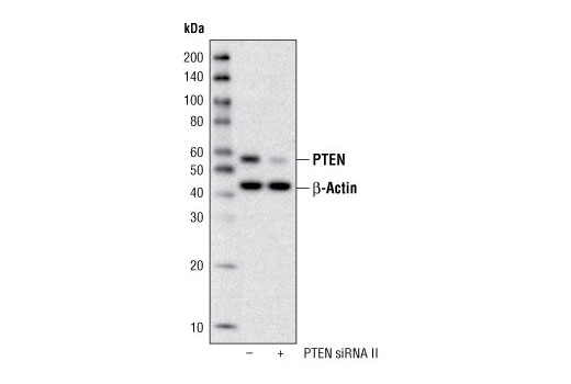
Western blot analysis of extracts from HeLa cells, transfected with 100 nM SignalSilence ® Control siRNA (Fluorescein Conjugate) #6201 (-) or SignalSilence ® PTEN siRNA II (+), using PTEN (138G6) Rabbit mAb and β-Actin (13E5) Rabbit mAb #4970. PTEN (138G6) Rabbit mAb confirms silencing of PTEN expression, while the β-Actin (13E5) Rabbit mAb is used to control for loading and specificity of PTEN siRNA.

Western blot analysis of extracts from A431, HeLa, 293, COS, PC12, NIH/3T3 and Mouse Brain cells, using PTEN (138G6) Rabbit mAb.

Immunohistochemical analysis of paraffin-embedded MDA-MB-468 xenograft, using Phospho-Akt (Ser473) (736E11) Rabbit mAb (IHC Preferred) (#3787) (left) or PTEN (138G6) Rabbit mAb (right). MDA-MB-468 cells lack PTEN. Note the lack of PTEN staining in the Phospho-Akt positive cells.

Immunohistochemical analysis of paraffin-embedded human lung carcinoma (left) and prostate carcinoma (right), using PTEN (138G6) Rabbit mAb. Note the stromal cell staining in the PTEN negative lung carcinoma, and the cancer cell staining in the PTEN positive prostate carcinoma.

Immunohistochemical analysis of paraffin-embedded human colon carcinoma, using PTEN (138G6) Rabbit mAb in the presence of control peptide (left) or PTEN Blocking Peptide #1250 (right).

Immunohistochemical analysis of paraffin-embedded cell pellets demonstrating the specificity of PTEN (138G6) Rabbit mAb: DU145, HT-29 and MCF-7 (PTEN positive) and Jurkat, MDA-MB-468 and LNCaP (PTEN negative).

Immunohistochemical analysis of paraffin-embedded xenografts using PTEN (138G6) Rabbit mAb. DU145 (left) and A549 (middle) are PTEN positive cell lines, while U-87MG (right) is PTEN negative.

Immunohistochemical analysis of paraffin-embedded MDA-MB-468 xenograft using Phospho-Akt (Ser473) (D9E) Rabbit mAb #4060 (left) or PTEN (138G6) Rabbit mAb (right). Note the presence of P-Akt staining in the PTEN deficient MDA-MB-468 cells.


