Formalin-fixed and paraffin-embedded human cancer tissue reacted with the primary antibody, which was peroxidase-conjugated to the secondary antibody, followed by DAB staining. This data demonstrates the use of this antibody for immunohistochemistry; clinical relevance has not been evaluated. BC = breast carcinoma; HC = hepatocarcinoma.

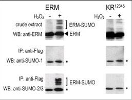
COS-7 cells were transfected for 24 hrs with a plasmid expressing FLAG-ERM (left panels) or FLAG-ERM KR12345 (right panels). Untreated (-) and H2O2-treated (+) cells were collected for immunoblot analysis. Top panels: cell lysates probed by western blot (WB) with an anti-ERM antibody. Center panels: cell lysates immunoprecipitated (IP) with an anti-FLAG antibody followed by WB SUMO-1 antibody. Bottom panels: cell lysates immunoprecipitated with an anti-FLAG antibody followed by WB with SUMO-2/3 antibody. (*) represents immunoprecipitated ERM-like forms recognized by anti-SUMO antibodies.

The anti-SUMO1 polyclonal antibody is used in Western blot to detect GST-SUMO1 fusion protein.

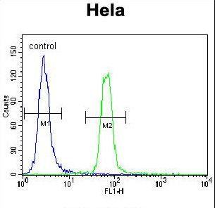
SUMO1 Antibody flow cytometry of HeLa cells (right histogram) compared to a negative control cell (left histogram). FITC-conjugated goat-anti-rabbit secondary antibodies were used for the analysis.


