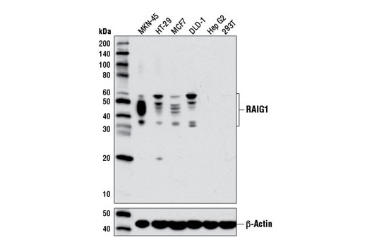
Western blot analysis of extracts from various cell lines using RAIG1 (D4S7D) XP ® Rabbit mAb (upper) or β-Actin (D6A8) Rabbit mAb #8547 (lower)
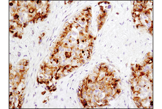
Immunohistochemical analysis of paraffin-embedded breast carcinoma using RAIG1 (D4S7D) XP ® Rabbit mAb.
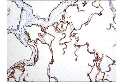
Immunohistochemical analysis of paraffin-embedded human normal lung using RAIG1 (D4S7D) XP ® Rabbit mAb.
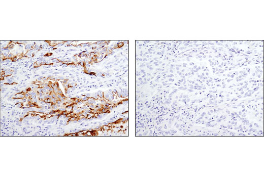
Immunohistochemical analysis of paraffin-embedded human lung carcinoma using RAIG1 (D4S7D) XP ® Rabbit mAb in the presence of control peptide (left) or antigen-specific peptide (right).
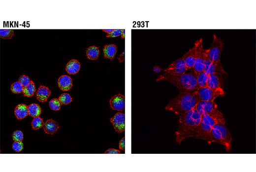
Confocal immunofluorescent analysis of MKN-45 (left) and 293T (right) cells using RAIG1 (D4S7D) XP ® Rabbit mAb (green). Actin filaments were labeled with DyLight® 554 Phalloidin #13054. Blue pseudocolor = DRAQ5 ® #4084 (fluorescent DNA dye).


