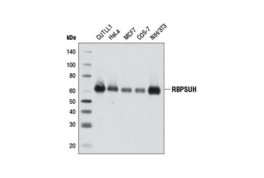
Western blot analysis of extracts from various cell lines using RBPSUH (D10A4) XP ® Rabbit mAb.

Immunohistochemical analysis of paraffin-embedded human colon carcinoma using RBPSUH (D10A4) XP ® Rabbit mAb.

Immunohistochemical analysis of paraffin-embedded human lung carcinoma using RBPSUH (D10A4) XP ® Rabbit mAb in the presence of control peptide (left) or antigen-specific peptide (right).

Immunohistochemical analysis of paraffin-embedded E18.5 mouse lung, Rbpjk F/+ Shh +/+ (wild type, left) or Rbpjk F/- Shh cre/+ (Rbpjk conditional knock out, right), using RBPSUH (D10A4) XP ® Rabbit mAb. Note lack of staining in the bronchial epithelial cells in the conditional knock out tissue (right). Tissue courtesy of Dr. Wellington Cardosa, Boston University School of Medicine.

Immunohistochemical analysis of paraffin-embedded human lung carcinoma using RBPSUH (D10A4) XP ® Rabbit mAb.

Immunohistochemical analysis of paraffin-embedded mouse lymph node using RBPSUH (D10A4) XP ® Rabbit mAb.

CUTLL1 cells were cultured in media with γ-secretase inhibitor (1 μM, 3 d) and then either harvested immediately (left panel) or washed and cultured in fresh media for 3 h (right panel). Chromatin immunoprecipitations were performed with cross-linked chromatin from 4 x 10 6 cells and 10 µl of RBPSUH (D10A4) XP ® Rabbit mAb or 2 µl of Normal Rabbit IgG #2729 using SimpleChIP ® Enzymatic Chromatin IP Kit (Magnetic Beads) #9003. The enriched DNA was quantified by real-time PCR using human HES1 promoter primers, SimpleChIP ® Human HES4 Promoter Primers #7273, and SimpleChIP ® Human α Satellite Repeat Primers #4486. The amount of immunoprecipitated DNA in each sample is represented as signal relative to the total amount of input chromatin, which is equivalent to one.


