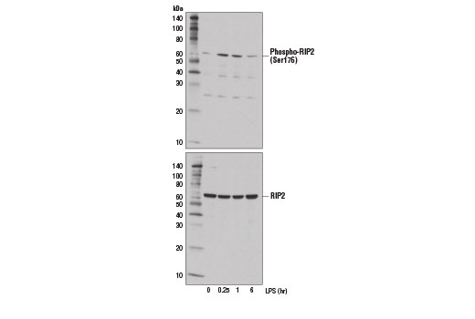
Western blot analysis of extracts from THP-1 cells, differentiated with TPA #4174 (80 nM, 24 hr) followed by treatment with LPS (1 μg/ml) for the indicated times, using Phospho-RIP2 (Ser176) (E1I9J) Rabbit mAb (upper) and RIP2 (D10B11) Rabbit mAb #4142 (lower).

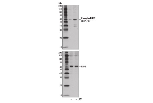
Western blot analysis of extracts from OVCAR8 cells, untreated (-) or UV-treated (+), using Phospho-RIP2 (Ser176) (E1I9J) Rabbit mAb (upper) and RIP2 (D10B11) Rabbit mAb #4142 (lower).

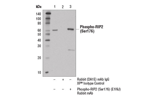
Immunoprecipitation of phospho-RIP2 (Ser176) from THP-1 cells, treated with TPA #4174 (80 nM, 24 hr) followed by LPS (1 μg/ml, 15 min), using Rabbit (DA1E) mAb IgG XP ® Isotype Control #3900 (lane 2) or Phospho-RIP2 (Ser176) (E1I9J) Rabbit mAb (lane 3). Lane 1 represents 10% input. Western blot was performed using Phospho-RIP2 (Ser176) (E1I9J) Rabbit mAb. Mouse Anti-rabbit IgG (Conformation Specific) (L27A9) mAb #3678 was used as a secondary antibody to avoid cross reactivity with rabbit IgG.


