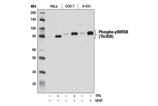
Western blot analysis of extracts from HeLa, COS-7, and A-431 cells, starved overnight and either untreated (-) or treated (+) with TPA #4174 (200 nM, 15 min) or Human Epidermal Growth Factor (hEGF) #8916 (100 ng/mL, 15 min), using Phospho-p90RSK (Thr359) (D1E9) Rabbit mAb.
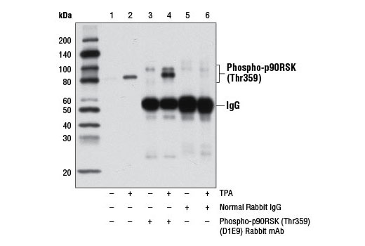
Immunoprecipitation of phospho-p90RSK (Thr359) from extracts of HeLa cells, serum starved overnight and either untreated (-) or treated with TPA #4174 (200 nM, 15 min; +), using Normal Rabbit IgG #2729 (lanes 5 and 6) or Phospho-p90RSK (Thr359) (D1E9) Rabbit mAb (lanes 3 and 4). Lanes 1 and 2 are 10% input. Western blot analysis was performed using RSK1/RSK2/RSK3 Antibody #9347.
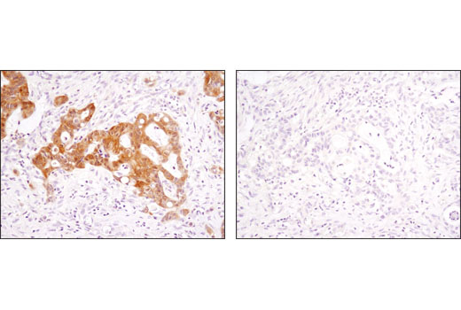
Immunohistochemical analysis of paraffin-embedded human ovarian carcinoma, control (left) or λ phosphatase-treated (right), using Phospho-p90RSK (Thr359) (D1E9) Rabbit mAb.
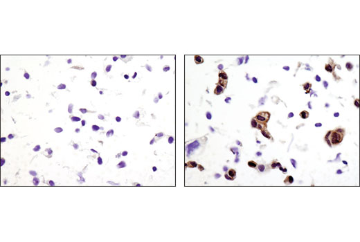
Immunohistochemical analysis of paraffin-embedded A-431 cell pellets, untreated (left) or treated with hEGF #8916 (right), using Phospho-p90RSK (Thr359) (D1E9) Rabbit mAb.
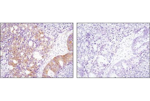
Immunohistochemical analysis of paraffin-embedded colon carcinoma using Phospho-p90RSK (Thr359) (D1E9) Rabbit mAb in the presence of control peptide (left) or antigen-specific peptide (right).
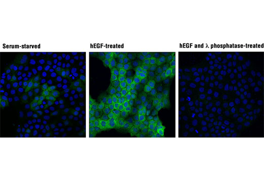
Confocal immunofluorescent analysis of A-431 cells, serum-starved (left), treated with hEGF #8916 (100 ng/mL, 15 min; center), or treated with hEGF and λ phosphatase (right), using Phospho-p90RSK (Thr359) (D1E9) Rabbit mAb (green). Blue pseudocolor = DRAQ5 ® #4084 (fluorescent DNA dye).