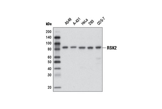
Western blot analysis of extracts from various cell lines using RSK2 (D21B2) XP ® Rabbit mAb.
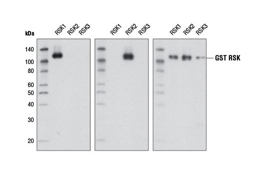
Western blot analysis of recombinant GST-tagged RSK1, RSK2, and RSK3 (2 μg each) using RSK1 (D6D5) Rabbit mAb #8408 (left panel), RSK2 (D21B2) XP ® Rabbit mAb #5528 (center panel), or GST (91G1) Rabbit mAb #2625 (right panel).
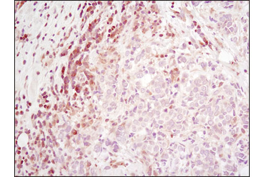
Immunohistochemical analysis of paraffin-embedded human breast carcinoma using RSK2 (D21B2) XP ® Rabbit mAb.
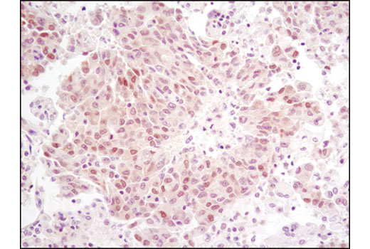
Immunohistochemical analysis of paraffin-embedded human lung carcinoma using RSK2 (D21B2) XP ® Rabbit mAb.
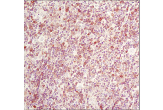
Immunohistochemical analysis of paraffin-embedded human lymphoma using RSK2 (D21B2) XP ® Rabbit mAb.
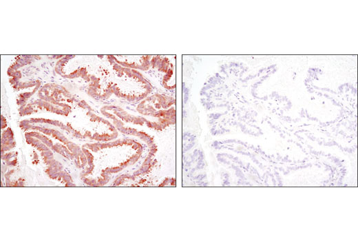
Immunohistochemical analysis of paraffin-embedded human ovarian serous adenocarcinoma using RSK2 (D21B2) XP ® Rabbit mAb in the presence of control peptide (left) or antigen-specific peptide (right).
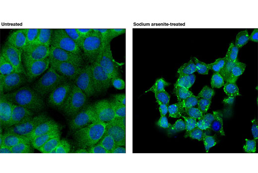
Confocal immunofluorescent analysis of MCF7 cells, untreated (left) or treated with sodium arsenite (500 uM for 1 hr; right) using RSK2 (D21B2) XP ® Rabbit mAb (green). Blue pseudocolor = DRAQ5 ® #4084 (fluorescent DNA dye). Localization of RSK2 to "stress granules" in arsenite-treated cells is very similar to that seen by Eisinger-Mathason et. al. (Molecular Cell, Volume 1, 5 September 2008, Pages 722-736).
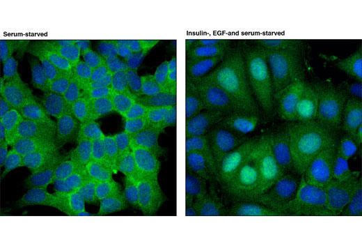
Confocal immunofluorescent analysis of MCF7 cells, untreated (left) or treated with insulin, EGF, and serum (right), using RSK2 (D21B2) XP ® Rabbit mAb (green). Blue pseudocolor = DRAQ5 ® #4084 (fluorescent DNA dye).
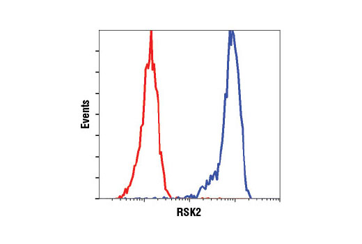
Flow cytometric analysis of HeLa cells using RSK2 (D21B2) XP ® Rabbit mAb (blue) compared to Rabbit (DA1E) mAb IgG XP ® Isotype Control #3900 (red).


