Neuropilin‑1 was detected in immersion fixed frozen sections of embryonic rat spinal cord (15 d.p.c.) using 5 µg/mL Goat Anti‑Rat Neuropilin‑1 Antigen Affinity-purified Polyclonal Antibody (Catalog # AF566) overnight at 4 °C. Tissue was stained with the Anti-Goat HRP-DAB Cell & Tissue Staining Kit (brown; Catalog # CTS008) and counterstained with hematoxylin (blue). View our protocol for Chromogenic IHC Staining of Frozen Tissue Sections.

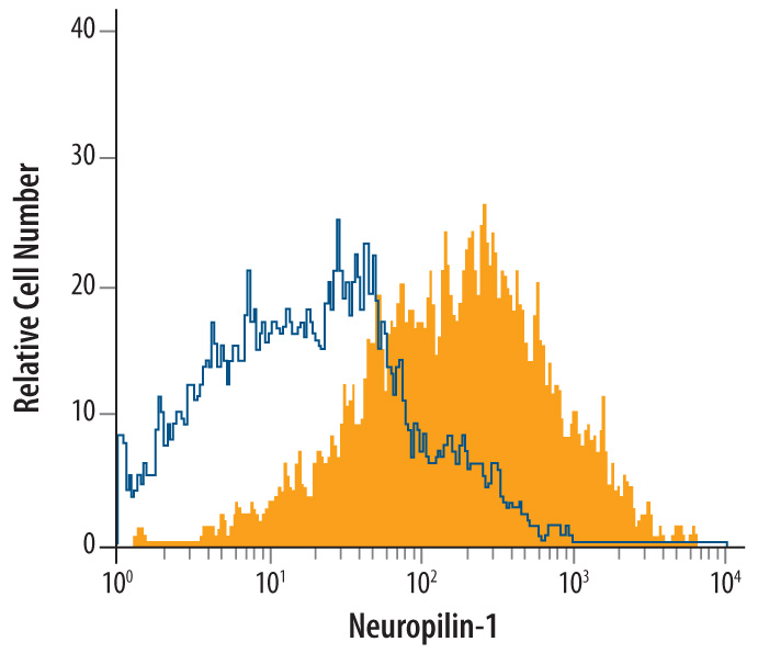
bEnd.3 mouse endothelioma cell line was stained with Goat Anti-Rat Neuropilin‑1 Antigen Affinity-purified Polyclonal Antibody (Catalog # AF566, filled histogram) or isotype control antibody (Catalog # AB‑108‑C, open histogram), followed by Allophycocyanin-conjugated Anti-Goat IgG Secondary Antibody (Catalog # F0108).

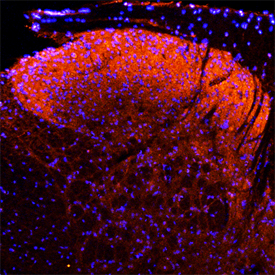
Neuropilin‑1 was detected in perfusion fixed frozen sections of rat spinal cord using Goat Anti-Rat Neuropilin‑1 Antigen Affinity-purified Polyclonal Antibody (Catalog # AF566) at 15 µg/mL overnight at 4 °C. Tissue was stained using the NorthernLights™ 557-conjugated Anti-Goat IgG Secondary Antibody (red; Catalog # NL001) and counterstained with DAPI (blue). Specific staining was localized to the dorsal horn. View our protocol for Fluorescent IHC Staining of Frozen Tissue Sections.


