
Recombinant Rat TNF‑ alpha (Catalog # 510-RT) induces cytotoxicity in the the L‑929 mouse fibroblast cell line in a dose-dependent manner (orange line), as measured by Resazurin. Cytotoxicity elicited by Recombinant Rat TNF‑ alpha (0.25 ng/mL) is neutralized (green line) by increasing concentrations of Goat Anti-Rat TNF‑ alpha Antigen Affinity-purified Polyclonal Antibody (Catalog # AF-510-NA). The ND50 is typically 0.15‑0.75 µg/mL in the presence of the metabolic inhibitor actinomycin D (1 µg/mL).
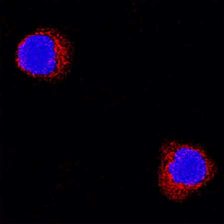
TNF‑ alpha was detected in immersion fixed rat splenocytes using Goat Anti-Rat TNF‑ alpha Antigen Affinity-purified Polyclonal Antibody(Catalog # AF-510-NA) at 15 µg/mL for 3 hours at room temperature. Cells were stained using the NorthernLights™ 557-conjugated Anti-Goat IgG Secondary Antibody (red; Catalog # NL001) and counterstained with DAPI (blue). Specific staining was localized to cytoplasmic. View our protocol for Fluorescent ICC Staining of Non-adherent Cells.