Recombinant Human Semaphorin 3E (Catalog # 3239-S3) inhibits proliferation in the HUVEC human umbilical vein endothelial cells in a dose-dependent manner (orange line). Plexin D1 mediated inhibition elicited by Recombinant Human Semaphorin 3E (5 ug/mL) is neutralized (green line) by increasing concentrations of Goat Anti-Human Plexin D1 Antigen Affinity-purified Polyclonal Antibody (Catalog # AF4160). The ND50 is typically 0.5-2 ug/mL.

Human peripheral blood monocytes were stained with Goat Anti-Human Plexin D1 Antigen Affinity‑purified Polyclonal Antibody (Catalog # AF4160, filled histogram) or control antibody (Catalog # AB‑108‑C, open histogram), followed by Phycoerythrin-conjugated Anti-Goat IgG Secondary Antibody (Catalog # F0107).

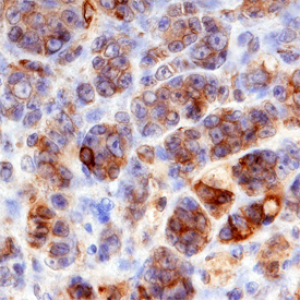
Plexin D1 was detected in immersion fixed paraffin-embedded sections of human melanoma using Goat Anti-Human Plexin D1 Antigen Affinity-purified Polyclonal Antibody (Catalog # AF4160) at 3 µg/mL overnight at 4 °C. Before incubation with the primary antibody, tissue was subjected to heat-induced epitope retrieval using Antigen Retrieval Reagent-Basic (Catalog # CTS013). Tissue was stained using the Anti-Goat HRP-DAB Cell & Tissue Staining Kit (brown; Catalog # CTS008) and counterstained with hematoxylin (blue). Specific staining was localized to plasma membranes. View our protocol for Chromogenic IHC Staining of Paraffin-embedded Tissue Sections.

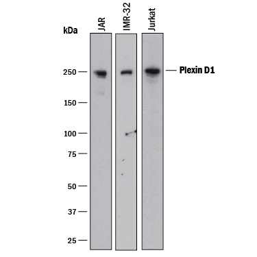
Western blot shows lysates of JAR human choriocarcinoma cell line, IMR‑32 human neuroblastoma cell line, and Jurkat human acute T cell leukemia cell line. PVDF membrane was probed with 0.5 µg/mL of Goat Anti-Human Plexin D1 Antigen Affinity-purified Polyclonal Antibody (Catalog # AF4160) followed by HRP-conjugated Anti-Goat IgG Secondary Antibody (Catalog # HAF017). A specific band was detected for Plexin D1 at approximately 250 kDa (as indicated). This experiment was conducted under reducing conditions and using Immunoblot Buffer Group 1.


