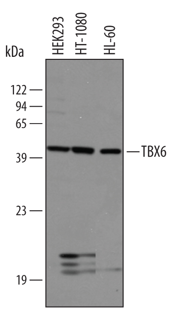
Western blot shows lysates of HEK293 human embryonic kidney cell line, HT1080 human fibrosarcoma cell line, and HL‑60 human acute promyelocytic leukemia cell line. PVDF membrane was probed with 1 µg/mL of Human TBX6 Antigen Affinity-purified Polyclonal Antibody (Catalog # AF4744) followed by HRP-conjugated Anti-Goat IgG Secondary Antibody (Catalog # HAF019). A specific band was detected for TBX6 at approximately 45 kDa (as indicated). This experiment was conducted under reducing conditions and using Immunoblot Buffer Group 8.

TBX6 was detected in immersion fixed frozen sections of embryonic mouse mesoderm (E9.5) using 10 µg/mL Human TBX6 Antigen Affinity-purified Polyclonal Antibody (Catalog # AF4744) overnight at 4 °C. Tissue was stained with the NorthernLights™ 557-conjugated Anti-Goat IgG Secondary Antibody (red; Catalog # NL001) and counterstained with DAPI (blue). View our protocol for Fluorescent IHC Staining of Frozen Tissue Sections.