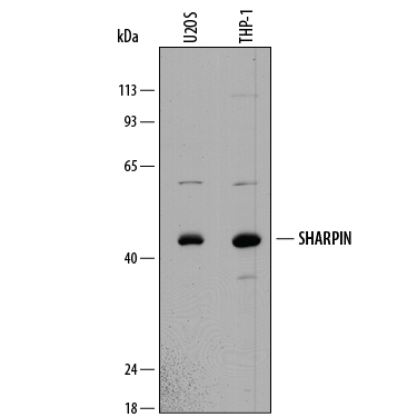
Western blot shows lysates of U2OS human osteosarcoma cell line and THP‑1 human acute monocytic leukemia cell line. PVDF membrane was probed with 1 µg/mL of Sheep Anti-Human SHARPIN Antigen Affinity-purified Polyclonal Antibody (Catalog # AF8100) followed by HRP-conjugated Anti-Sheep IgG Secondary Antibody (Catalog # HAF016). A specific band was detected for SHARPIN at approximately 43 kDa (as indicated). This experiment was conducted under reducing conditions and using Immunoblot Buffer Group 1.
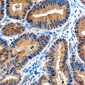
SHARPIN was detected in immersion fixed paraffin-embedded sections of human ovary using Sheep Anti-Human SHARPIN Antigen Affinity-purified Polyclonal Antibody (Catalog # AF8100) at 10 µg/mL overnight at 4 °C. Tissue was stained using the Anti-Sheep HRP-DAB Cell & Tissue Staining Kit (brown; Catalog # CTS019) and counterstained with hematoxylin (blue). Specific staining was localized to cytoplasm. View our protocol for Chromogenic IHC Staining of Paraffin-embedded Tissue Sections.