
Western blot shows lysates of BG01V human embryonic stem cells. PVDF membrane was probed with 1 µg/mL of Mouse Anti-Human ZNF281 Monoclonal Antibody (Catalog # MAB6350) followed by HRP-conjugated Anti-Mouse IgG Secondary Antibody (Catalog # HAF018). A specific band was detected for ZNF281 at approximately 100 kDa (as indicated). This experiment was conducted under reducing conditions and using Immunoblot Buffer Group 1.

ZNF281 was detected in immersion fixed BG01V human embryonic stem cells using Mouse Anti-Human ZNF281 Monoclonal Antibody (Catalog # MAB6350) at 10 µg/mL for 3 hours at room temperature. Cells were stained using the NorthernLights™ 557-conjugated Anti-Mouse IgG Secondary Antibody (red, upper panel; Catalog # NL007) and counterstained with DAPI (blue, lower panel). Specific staining was localized to nuclei. View our protocol for Fluorescent ICC Staining of Stem Cells on Coverslips.
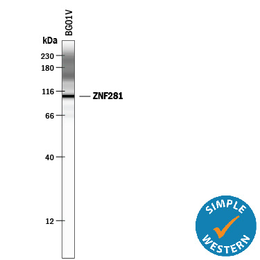
Simple Western lane view shows lysates of BG01V human embryonic stem cells, loaded at 0.5 mg/mL. A specific band was detected for ZNF281 at approximately 107 kDa (as indicated) using 50 µg/mL of Mouse Anti-Human ZNF281 Monoclonal Antibody (Catalog # MAB6350). This experiment was conducted under reducing conditions and using the 12-230 kDa separation system. Non-specific interaction with the 230 kDa Simple Western standard may be seen with this antibody.