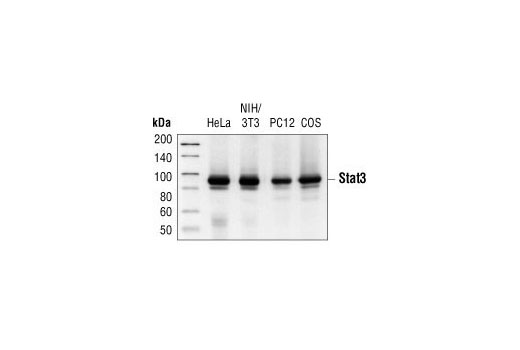
Western blot analysis of extracts from HeLa, NIH/3T3, PC12 and COS cells, using Stat3 (124H6) Mouse mAb.

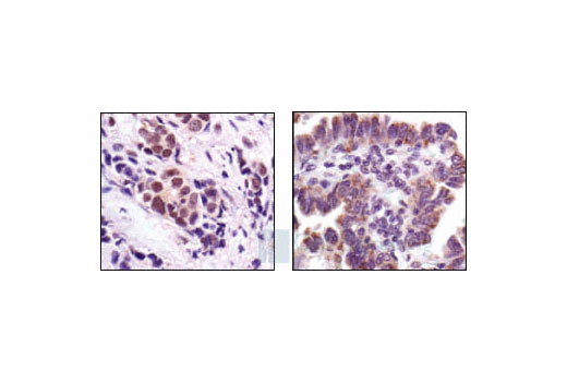
Immunohistochemical analysis of paraffin-embedded human breast carcinoma (left), showing nuclear and cytoplasmic staining, and human lung carcinoma (right), showing cytoplasmic staining, using Stat3 (124H6) Mouse mAb.

Confocal immunofluorescent analysis of HeLa cells either serum-starved (left) or IFNalpha-treated (right) and labeled with Stat3 (124H6) Mouse mAb (green).

Flow cytometric analysis of Hela cells, untreated (blue) or IFN-alpha-treated (green), using Stat3 (124H6) Mouse mAb compared with a nonspecific negative control antibody (red).

Chromatin immunoprecipitations were performed with cross-linked chromatin from 4 x 10 6 Hep G2 cells starved overnight and treated with IL-6 (100 ng/ml) for 30 minutes, and either 10 μl of Stat3 (124H6) Mouse mAb or 2 μl of Normal Rabbit IgG #2729 using SimpleChIP ® Enzymatic Chromatin IP Kit (Magnetic Beads) #9003. The enriched DNA was quantified by real-time PCR using human IRF-1 promoter primers, SimpleChIP ® Human c-Fos Promoter Primers #4663, and SimpleChIP ® Human α Satellite Repeat Primers #4486. The amount of immunoprecipitated DNA in each sample is represented as signal relative to the total amount of input chromatin, which is equivalent to one.


