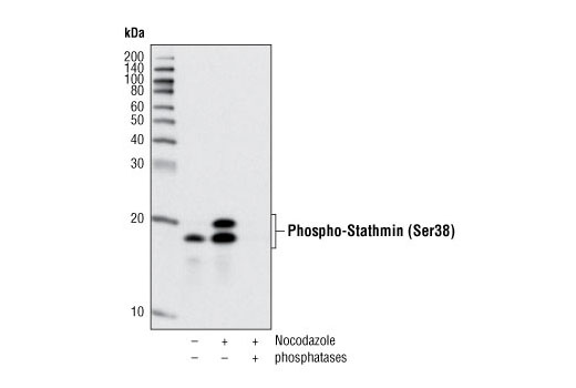
Western blot analysis of extracts from HT-29 cells, untreated or treated with nocodazole alone (100 ng/mL 24 hours) or nocodazole followed by λ and calf intestinal phosphatases, using Phospho-Stathmin (Ser38) (D19H10) Rabbit mAb.

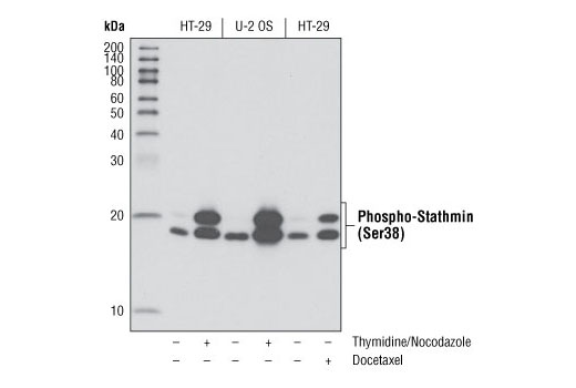
Western blot analysis of extracts from HT-29 and U-2 OS cells, untreated or synchronized in mitosis, using Phospho-Stathmin (Ser38) (D19H10) Rabbit mAb. Mitotic synchrony was performed by using either a thymidine block followed by release into 100 ng/mL nocodazole for 24 hours or using 100 ng/mL Docetaxel #9886 for 24 hours.

Immunohistochemical analysis of paraffin-embedded human breast carcinoma control (left) or λ phosphatase-treated (right) using Phospho-Stathmin (Ser38) (D19H10) Rabbit mAb.

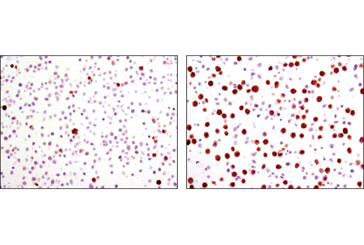
Immunohistochemical analysis of paraffin-embedded HT-29 cell pellets, untreated (left) or nocodazole-treated (right), using Phospho-Stathmin (Ser38) (D19H10) Rabbit mAb.

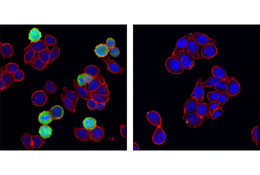
Confocal immunofluorescent analysis of HT-29 cells, untreated (left) or λ phosphatase-treated (right), using Phospho-Stathmin (Ser38) (D19H10) Rabbit mAb (green). Actin filaments were labeled with DY-554 phalloidin (red). Blue pseudocolor = DRAQ5 ® #4084 (fluorescent DNA dye).


